Translate this page into:
Vertical incision subperiosteal tunnel access and three-dimensional OBS lever arm to recover a labially-impacted canine: Differential biomechanics to control root resorption
First author: Dr. Jia Hong Lin,

*Corresponding author: Dr. Roberts W. Eugene, Department of Emeritus of Orthodontics, Indiana University, School of Dentistry, 1121 W. Michigan St. Indianapolis, IN 46202, USA. werobert@iu.edu, werobert@me.com
-
Received: ,
Accepted: ,
Abstract
A 15-year-old female presented with a chief complaint of unesthetic smile and protrusive lips. Lower facial height and convexity were within normal limits, but the lower lip was protrusive (3mm to the E-Line). Bimaxillary retrusion (SNA 79.5˚, SNB 76˚, and ANB 3.5˚) and a high mandibular angle (SN-MP 38˚) were noted. Lower incisors were prominent (L1 to MP 96˚ and L1 to NB 8 mm). Molars were Class I, but the upper right canine (UR3) was Class II. The upper left deciduous canine (ULc) was retained, and the UL3 was labially impacted. An oblique direction of canine eruption wedged the impaction between the keratinized mucosa and the adjacent incisor, eliciting root resorption on the labial surface of the UL2. The discrepancy index (DI) was 16. Following extraction of all four first premolars and the ULc, all teeth except the UL2 were bonded with a Damon Q® passive self-ligating bracket system. Vertical incision subperiosteal tunnel access (VISTA) technique was performed to produce a submucosal space for retraction and extrusion of the impacted UR3. A button was bonded on the UL3, and a power chain was attached. The elastomer chain exited the mucosa through a more distal incision, and traction was applied with a custom lever arm, anchored by an OBS® inserted into the left infrazygomatic crest. The impaction was retracted into a normal position between the UL2 and UL4. Once the UL3 was extruded to the occlusal plane, the UL2 was bonded and its axial inclination was corrected with a labial root torquing auxiliary. Both arches were detailed and finished. After 24 months of active treatment, the UL3 was well aligned, but the labial gingiva supporting it was immature and only partially keratinized. Follow-up visit 1.5 years later showed its maturation into a stable but relatively thin band of gingiva. In retrospect, this UL3 gingival problem may have been avoided by adjusting the three-dimensional (3D) lever arm for a more palatal emersion of the impaction. There was no change in the preexisting labial root resorption of the UL2, but no additional root resorption on any teeth occurred during active treatment. Final alignment and dental esthetics were excellent as evidenced by an American Board of Orthodontics Cast-Radiograph Evaluation score of 12, and the IBOI Pink and White Esthetic Score of 2. VISTA with an OBS 3D lever arm is an important advance for orthodontic impaction recovery. Submucosal retraction of a labially- impacted, partially transposed maxillary canine permits optimal emergence into the arch. Differential biomechanics of soft and hard tissue explains impaction-related root loss before treatment, as well as the mechanism for protecting an unrestrained lateral incisor while the impacted canine is recovered.
Keywords
Bone screw anchorage
Dental sac
Differential biomechanics
Eruptive force
Follicle
Impacted maxillary canine
Root resorption
Tooth movement
Vertical incision subperiosteal tunnel access
INTRODUCTION
Dental nomenclature for this report is a modified Palmer notation with four oral quadrants: Upper right (UR), upper left (UL), lower right (LR), and lower left (LL). From the midline permanent teeth are numbered 1-8, and deciduous teeth are delineated a-e. Management of impacted maxillary canines (U3s) is one of the most challenging tasks for orthodontists. Studies have shown a prevalence of 0.27–2.4%,[1,2] second only to third molars.[3] In North American patients, about two-thirds of the impacted canines are located palatally, with the rest positioned labially or within the alveolus.[4] In contrast, ethnic Chinese adolescents experience 49.85–67.7% of impacted canines on the labial side.[5,6] Labial impactions are more difficult to manage clinically because the recovery process is prone to root resorption and gingival recession.[7-9]
For labial impactions above the mucogingival junction (MGJ), Kokich[10] proposed the apically positioned flap (APF) or the closed eruption (CE) technique. The latter is favored because it does not expose the roots of the adjacent lateral incisors, which may result in devitalization.[8,11] Furthermore, it decreases the possibility of re-intrusion and gingival scarring.[12] Loss of attachment and gingival recession are best controlled with the tissue tunneling approach introduced by Crescini et al.[13]
Closed flap surgical approaches are well established for managing impactions in the maxillary anterior esthetic zone,[14] but an impacted U3s with mesial transposition into the adjacent lateral incisor is a particularly challenging problem, both with respect to mechanics and preservation of gingival health. Traction of the impaction through the center of the alveolar ridge may impinge particularly on the adjacent lateral incisor, resulting in slow movement, and/or extensive root resorption.[15] To avoid these problems, Su et al.[16] modified the Zadeh[17] vertical incision subperiosteal tunnel access (VISTA) technique to preserve gingival margins. Mesially-displaced, impacted U3s are retracted and extruded within the submucosal space. This minimally- invasive approach permits movement of the impaction away from adjacent teeth; it is then positioned vertically in the arch before emerging through the mucosa.[9]
HISTORY AND ETIOLOGY
A relatively immature 15-year-old 4 months female sought orthodontic consultation for unesthetic maxillary anterior dentition and protrusive lips [Figure 1]. No contributing medical or dental history was reported, but some late facial growth was expected. Clinical examination revealed a convex facial profile and lip protrusion that was slightly protrusive, particularly to the ideal Chinese standard.[18] Overbite and overjet of the central incisors were within normal limits (WNL), and the buccal segments were Class I, but there was a bilateral irregularity in the maxillary lateral incisor and canine region [Figures 2 and 3]. An edge-to-edge relationship was noted between the UR and LR lateral incisors, UR2 and LR2, respectively. Maximal overjet was 4 mm for the upper left lateral incisor (UL2). The deciduous ULc was retained with no mobility. Crowding was about 6 mm in the upper and 4 mm in the lower arches. Panoramic [Figure 4] and lateral cephalometric [Figure 5] radiographs revealed impaction of the UL3. Cone beam computed tomography (CBCT) images [Figures 6 and 7] showed that the impacted UL3: (1) Was impacted on the labial surface, (2) Had a mesially and labially inclined crown, and (3) Was impinged on the labial surface of the UL2 root. The root of the ULc was not resorbed, but modest root resorption was noted on the labial aspect of the apical half of the UL2 root [Figure 7].

- Pre-treatment facial and intraoral photographs.

- Pre-treatment upper left deciduous canine associated with a mesially and labially displaced UL2 crown.

- Pre-treatment dental models (casts).

- Pre-treatment panoramic radiograph.

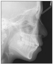
- Pre-treatment lateral cephalometric radiograph.

- Cone beam computed tomography image of the maxillary dentition shows a labially-positioned impacted UL3 over the root of UL2.

- Cone beam computed tomography cut through the long axis of the UL2 shows labial impingement of the impacted UL3 (arrow). Compression of the interposed soft tissues (dental sac and periodontal ligament) results in damage to the tooth root which is followed by resorption.
DIAGNOSIS
Facial
Convexity: WNL (12˚)
Lip protrusion: Slightly protrusive (0 mm/3 mm to the E-line).
Skeletal
Sagittal Relationship: Bimaxillary retrusion (SNA 79.5˚, SNB 76˚, and ANB 3.5˚)
Mandibular Plane Angle: Increased (SN-MP 38˚ and FMA 31˚).
Dental
Occlusion: Class I molar
Overjet: 4 mm
Lower incisor: Protrusive (L1-NB 8mm), increased axial inclination (L1-MP 96˚)
Impaction: Labially impacted UL3, crown transposed impinging on the UL2 root, American Board of Orthodontics DI: 16.
TREATMENT OBJECTIVES
Maxilla and mandible - normal growth expression in A-P, vertical, and transverse planes
Maxillary dentition
A - P: Retract incisors
Vertical: Maintain
Intercanine width: Decrease
Intermolar width: Decrease as molars are protracted to close L4 spaces.
Mandibular dentition
A - P: Retract incisors
Vertical: Allow extrusion consistent with normal growth
Inte-canine width: Maintain
Decrease as molars are protracted to close U4 spaces.
Facial esthetics
Lip Retraction: Retract upper and lower lips according to the ethnic preference.[18]
TREATMENT PLAN
Objectives for full fixed appliance treatment were to recover the impacted UL3, align the dentition, and retract the lips. Three options were considered:
Extract all four 1st premolars and the ULc. Use the modified VISTA and OBS three-dimensional (3D) lever arm technique to align the impacted UL3.
Extract UR4, LL4, LR4, ULc, and the impacted UL3. Substitute UL3 with UL4.
Extract only the deciduous canine. Use the modified VISTA and OBS 3D lever arm technique to align the impacted UL3.
First option
Extraction of premolars permits retraction of the lips, but specialized surgery and mechanics are required to recover the impacted canine. This approach was expected to have the longest treatment duration.
Second option
Premolars and the deciduous canine are extracted to achieve the patient’s desire for less lip protrusion. Extracting the impaction rather than recovering it would decrease treatment time, but substituting the UL4 for the missing UL3 results in an esthetic and functional compromise.
Third option
Extract only the ULc and recover the impacted UL3. This non-extraction approach offers the shortest treatment duration. Good dental esthetics and function are expected, but this plan is unlikely to correct lip protrusion.
After a thorough discussion of all three options, the patient and her parents preferred the first option because it delivered the most ideal dental and facial result, consistent with the family’s preferred ethnic standard.[18]
TREATMENT PROGRESS
Extraction of all four first premolars and the ULc was the first step in active treatment. A passive self-ligating (PSL) a fixed appliance (Damon Q®, Ormco Corporation, Glendora, CA) was bonded on all upper teeth except for the UL2, and a 0.014-in CuNiTi archwire was engaged. High-torque brackets were chosen for the two upper incisors to control a loss of torque (decreased axial inclination) during space closure. Not bonding the UL2 before UL3 recovery is a very important aspect of patient management. When the infringed tooth (UL2) is not engaged on the fixed appliance, it is free to move spontaneously out of the path of movement as the impact is recovered.[19]
When the crown of the impacted canine is positioned at or near the MGJ, it may spontaneously erupt into a high position much like the UR3. The initial treatment was planned with that possibility in mind. The first phase was to align all erupted teeth in the upper and lower arches, except the UL2. The archwire sequence was: 1. 0.014-in CuNiTi, 2. 0.014 × 0.025-in CuNiTi, and 0.017 × 0.025-in TMA. During the initial alignment phase, the impacted UL3 failed to erupt, and a panoramic radiograph 8 months into treatment showed no change in the position of the impaction, so surgical intervention was indicated.
The preferred surgical approach [Figure 8] was the VISTA technique of Zadeh,[17] as modified by Su et al.,[16] combined with infrazygomatic crest (IZC) OBS anchorage[19] and 3D lever arm mechanics [Figure 9].[20] CBCT imaging [Figurers 6 and 7] showed the precise location of the impaction, so the initial vertical incision was performed between the central and lateral incisors to expose the crown of the impaction [Figure 10a]. A periosteal elevator was then used to detach the periosteum and expose the UL3 [Figure 10b]. Bone covering the crown was removed down to the cementoenamel junction. The impacted canine was carefully luxated with an elevator to control for ankylosis, and then a button was bonded in the center of the exposed enamel. A power chain was attached to the button, a second vertical incision was made in the vestibule superior to the edentulous space, superior to the normal position of the UL3, and the power chain exited the submucosal tunnel [Figure 10c]. Subperiosteal decortication, of the alveolar bone surface in the path of UL3 retraction, was achieved with a#4 round carbide bur. An OBS® (iNewton Dental Ltd., Hsinchu City, Taiwan) was inserted to the left IZC, and a 3D lever arm was inserted into the rectangular hole of the anchorage device [Figure 9]. Finally, the power chain that was attached to the impaction delivered a distal traction force through the lever arm anchored by the IZC OBS. Following activation of the mechanism, the two vertical incisions were sutured to ensure minimal damage to the mucosa [Figures 10-12].

- The vertical incision subperiosteal tunnel access procedure is a novel, submucosal tunneling procedure originally designed to surgically correct gingival recession (a). Through vertical incisions, the labial mucosa is undermined and repositioned coronally as shown by the yellow arrow (b). The submucosal space fills with a hematoma (red) that provides platelet-derived growth factors to promote healing (c). This minimally invasive approach is utilized to correct soft tissue defects in the maxillary anterior region.

- A diagram superimposed on an intraoral photograph illustrates the design of the implant recovery mechanism in the sagittal plane. The UL3 impacted against the UL2 root is accessed with a vertical incision subperiosteal tunnel access vertical incision, and a button is bonded on the labial surface. A blue chain of elastics applies distal and occlusal traction to the UL3, through a three-dimensional lever arm inserted into the hole on an IZC OBS.

- (a) The first incision was made in the mucosa covering the crown of the impacted canine. (b) Periosteal elevators were used to reflect the incision and expose the crown for bonding the button. (c) A second incision was then made at the site where the power chain exits the soft tissue (arrow).

- (a) An OBS (white arrow) was inserted in the IZC to anchor the three-dimensional (3D) lever arm. (b) The distal end of the 3D lever arm was inserted in the hole of the OBS (green arrow). (c) The power chain attached to the UL3 was activated by the 3D lever arm in the direction of the yellow arrow.

- (a) The two incisions were then sutured for primary healing. (b) The occlusal view of the lever arm shows it was contoured away from the cheek to prevent soft tissue irritation. (c) The buccal view of the mechanics is illustrated with a drawing superimposed on the post-operative photograph. Red lines show 1st and 2nd sutured incisions and a gold chain of elastics show the line of traction. Note both ends of the lever-arm are secured with bonded resin.
Post-operative panoramic radiographs monitored the movement of the impacted canine relative to adjacent teeth [Figure 13]. After 7 months of activation, the UL3 was uprighted and internally positioned in the arch, coronal to the MGJ. The canine crown and button were visible beneath the transparent gingiva [Figure 14]. After 9 months of retraction, the canine erupted to the level of the occlusal plane, but its buccal gingiva was immature and bright red in color [Figure 15]. The crown of the UL3 was tipped to the buccal and rotated distal in relative to the adjacent premolar. A high torque PSL bracket was bonded on the UL3, and a standard torque bracket was bonded on the UL2 [Figure 15]. A light force, continuous archwire (0.014-in CuNiTi) was utilized to align the upper arch [Figure 16]. A sequence of three additional upper archwires (0.014 × 0.025-in CuNiTi, 0.017 × 0.025- in TMA, and 0.016 × 0.022-in SS) were used to refine the alignment [Figures 16 and 17]. Labial root torque was applied to the UL2 with a torquing auxiliary [Figure 18]. In the past month of treatment, the archwire was sectioned distal to the upper canines, and intermaxillary elastics (Chipmunk 1/8-in 3.5-oz, Ormco, Glendora, CA) were used for final finishing of the buccal segments [Figure 19].

- A panel of four radiographs shows progress in the recovery of the impacted UL3. Each radiograph is labeled with a code designating the time in months since vertical incision subperiosteal tunnel access surgery and initiation of traction (first number), and the number of months into active treatment (second number). Thus, the upper left view (0/8) is the immediate post-operative radiograph for the surgery performed at 8 months into treatment. The lower right image (5/13) shows the position of the UL3 after 5 months of traction, which corresponds to the 13 months of treatment.

- Left: After 15 months of active treatment including 7 months of traction (7/15), UL3 is correctly positioned in the sagittal plane, and there are no obstructions for extrusions. Right: The UL3 crown is visible underneath the overlying gingiva, which is immediately coronal to the mucogingival junction (white scalloped line). Note the line of traction for the lever arm is buccal and occlusal.

- Left: After 9 months of traction and 17 months of active treatment (9/17), the UL3 is extruded to the occlusal plane. Right: Brackets were bonded on the UL2 and UL3, and a CuNiTi archwire is used to align the arch. Note the large red area of immature, nonkeratinized gingiva (white arrow) which will mature into the band of keratinized gingiva supporting the UL3.

- Treatment progress for the upper arch is shown in months (M), and the archwire progression is specified from the start of treatment (0M) to 24 months (24M).

- Treatment progress for the lower arch is shown in months (M), and the archwire progression is specified from the start of treatment (0M) to 24 months (24M).

- At 15 months after surgery and 23 months into treatment (15/23), an auxiliary torquing spring (yellow arrow) is shown on the UL2 to torque the root labially.

- To finish the occlusal contacts in the buccal segments, the upper archwire is sectioned distal to the canines, and vertical elastics are applied as shown.
Following 25 months of active treatment, all brackets were removed and fixed retainers were constructed on the maxillary incisors (UR2-UL2) and the mandibular anterior segment (LR3-LL3). Maxillary anterior frenectomy and gingivectomy were performed with a diode laser to optimize dental esthetics [Figure 20]. Figure 21 is a panel of radiographs and photographs documenting the pre-treatment condition and the post-treatment outcome. The labial gingiva for the UL3 was irregular and only partially keratinized. For comparison, a 1.5-year follow-up view of the same region shows a narrow band of mature gingiva supporting the recovered UL3 [Figure 22].

- Following the removal of fixed appliances at 17 months after surgery, and 25 months into treatment (17/25), gingivectomy and frenectomy were performed in the maxillary anterior segment to enhance esthetics.

- Four illustrations show a coordinated radiograph and intraoral photograph of the pretreatment (Initial) condition in the two upper views, and the corresponding final records are shown in the lower panel (Final).

- At 1.5-year follow-up an intraoral photograph shows the relatively thin band of gingiva on the UL3 compared to adjacent teeth. Compare this follow-up view to Figures 15 and 27 to assess the maturation of the gingiva on the buccal surface of the UL3.
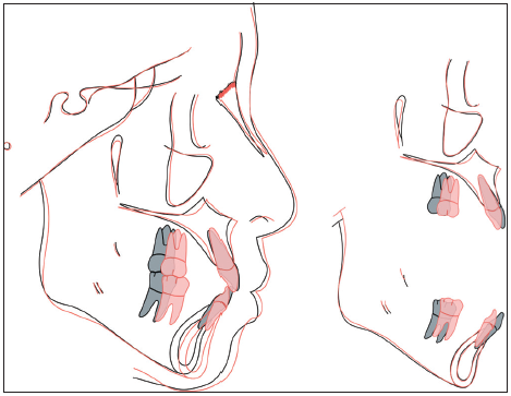
Post-treatment panoramic [Figure 23], model casts [Figure 24], and lateral cephalometric [Figure 25] radiographs document the outcome following 25 months of active surgical and orthodontic therapy. Superimposition of before and after the treatment cephalometric tracings shows the late growth and dentofacial orthopedic changes associated with active treatment [Figure 26].

- Post-treatment panoramic radiograph.

- Post-treatment dental models (casts) radiograph.

- Post-treatment lateral cephalometric radiograph.

- Initial (black) and final (red) cephalometric tracings are superimposed on the anterior cranial base (left) and the skeletal structures of the maxilla and the mandible (right).
RESULTS ACHIEVED
Maxilla (all three planes)
A - P: Increased
Vertical: Maintained
Transverse: Maintained.
Mandible (all three planes)
A - P: Increased
Vertical: Increased
Transverse: Maintained.
Maxillary dentition
A - P: Retraction of incisors, protraction of molars
Vertical: Maintained
Intermolar width: Maintained.
Mandibular dentition
A - P: Retraction of incisors, protraction of molars
Vertical: Slightly extruded consistent with normal growth
Inter-molar/Intercanine width: Decreased/Maintained.
Facial esthetics
Convexity: Decreased
Lips: Retraction of the upper and lower lips.
FINAL EVALUATION OF TREATMENT
Clinical examination revealed an improved facial profile, i.e., the nasomaxillary complex grew anteriorly as the lips were retracted [Figures 26 and 27]. Maxillary and mandibular incisors were retracted and uprighted, as evidenced by the decreased protrusion and axial inclination [Table 1]. The score for the Cast-Radiograph Evaluation was 17 points. The major discrepancy was axial inclinations in the final panoramic radiograph [Figure 23].

- Post-treatment facial and intraoral photographs.
| Regional measurements | Pre-TX | Post-TX | Diff. |
|---|---|---|---|
| Skeletal analysis | |||
| SNA˚ (82˚) | 79.5˚ | 81˚ | 1.5˚ |
| SNB˚ (80˚) | 76˚ | 78˚ | 2˚ |
| ANB˚ (2˚) | 3.5˚ | 3˚ | 0.5˚ |
| SN-MP˚ (32˚) | 38˚ | 37˚ | 1˚ |
| FMA˚ (25˚) | 31˚ | 30˚ | 1˚ |
| Dental analysis | |||
| U1 TO NA mm (4 mm) | 5 mm | 2 mm | 3 mm |
| U1 TO SN˚ (104˚) | 106˚ | 102˚ | 4˚ |
| L1 TO NB mm (4 mm) | 8 mm | 3 mm | 5 mm |
| L1 TO MP˚ (90˚) | 96˚ | 86.5˚ | 9.5˚ |
| Facial analysis | |||
| E-LINE UL (-1 mm) | 0 mm | 0 mm | 0 mm |
| E-LINE LL (0 mm) | 3 mm | 1.5 mm | 1.5 mm |
| %FH: Na-ANS-Gn (53%) | 53% | 53% | 0% |
| Convexity: G-Sn-Pg’ (13˚) | 12˚ | 13.5˚ | 1.5 |
The total treatment time was 25 months for the partially transposed labially impacted maxillary canine, which is similar to the only other comparative report in literature.[9] Post-treatment facial and intraoral photographs [Figure 27], as well as similar records at 1.5-year follow-up, showed that the recovered canine and adjacent lateral incisor (UL2) were stable. No signs of re-intrusion, significant root resorption, or inflammation of the soft tissue were noted. The keratinized gingiva around the UL3 was acceptable [Figure 22], but should be followed long term. Third molars were recommended for extraction [Figure 23].
DISCUSSION
VISTA[17] is a novel method for the management of labially-impacted canines.[16] The method as revised by Su et al.[19] preserves adequate keratinized tissue when the impaction emerges [Figure 22]. As the impaction is recovered, it is important to delay the bonding of the adjacent lateral incisor to control root resorption.[9] Pre-existing root loss does not recover, but it also does not progress if the impingement is carefully corrected as the impaction is retracted. The use of the 3D lever arm anchored by an IZC OBS is particularly useful. It can be adjusted for staged movement in all planes of space as needed.[20]
LABIAL IMPACTION EXPOSURE
A challenging aspect for recovery of labial impactions is maintaining keratinized gingival support. A minimum of 2 mm of keratinized gingiva is necessary to maintain gingival health.[21] Labial impactions may emerge through alveolar mucosa rather than keratinized gingiva, so some degree of long-term gingival compromise is common.[10-12] The VISTA procedure allows for submucosal movement of a transposed impaction to its correct position in the arch [Figure 13], before emergence through keratinized gingiva [Figure 21]. In retrospect, a wider band of keratinized tissue on the UL3 may have been possible with a more vertical vector of traction when the UL3 was extruded [Figure 14].
A crucial factor is the site of emergence relative to the MGJ. A frequently cited study by Kokich[10] laid out three options: Excisional uncovering (EU), APF, and CE. EU is applicable if the crown of the impaction is coronal to the MGJ, but both APF and CE are used for impactions positioned superior to the MGJ. Vermette et al.[12] reported that the CE approach was superior to APF because it was less susceptible to gingival scarring and recession. These problems with gingival healing are attributed to “overstretching” of the keratinized layer following the primary healing of the gingival attachment. When an exposed tooth is moved coronally, the mucosa stretching may exceed the proliferative potential of the tissue. Furthermore, the strain may be in an oblique direction that tends to asymmetrically retract the gingival margin. Exposing an impaction, and repositioning the keratinized tissue for a centimeter or more, may devitalize or compromise the periodontal support of an adjacent tooth. For labially-impacted maxillary anterior teeth, CE is more reliable than ARF for optimal esthetic outcomes.[10,12,14]
Crescini et al.[13] proposed a CE approach mimicking a natural eruption route through the middle of the alveolus by performing a tunneling procedure from the crown of the impaction to the socket of its extracted predecessor. A gold chain is bonded to the enamel of the impaction to permit traction along the prepared path. The average time elapsed between the application of traction and the emergence of the cusp of the impacted canine is 11 months. A 3-year follow-up study of the procedure showed no attachment loss or gingival recession. The problem with this approach is the requirement of no obstacles other than bone between the crown of the impaction and the desired emergence site. Partially or fully transposed teeth with an unfavorable orientation have a poor prognosis because the surgically prepared path would damage roots of adjacent teeth.[6,9,19] For the present patient, the preferred method was to retract the impacted canine away from the lateral incisor root with the OBS-anchored 3D lever arm to expedite the recovery without precipitating additional root resorption.[22]
Previous impact recovery methods have relied on variations of linear traction. Unfavorable position and transposition of impactions may require staged movement in multiple directions with differential loads for an optimal outcome with minimal collateral damage. The present patient required swinging the impaction around the root of the lateral incisor without penetrating the oral mucosa at the corner of the arch, i.e., canine eminence. For precise submucosal movement, Su et al.[16] proposed a modification of the Zadeh[17] VISTA procedure to retract an impaction with a 3D lever arm with modification of the line of traction, as needed [Figures 9, 11, 12, and 14]. To accelerate tooth movement, decortication was performed along the proposed traction path, a procedure referred to as the periodontally accelerated osteogenic orthodontics.[23] Through the VISTA and OBS 3D lever arm approach, the partially transposed UL3 was retracted and extruded to emerge in its correct position in 9 months [Figures 14 and 15].
DELAYED BONDING OF THE LATERAL INCISOR
When lateral incisors are not bracketed, and restrained by an archwire or other retaining device, they are free to move away from the encroachment of a tooth follicle.[9] Broadbent[24] described the guidance of eruption theory that is commonly deemed the “ugly duckling stage” to explain the crown flaring and/or mesial root movement of maxillary lateral incisors due to the development of the unerupted canines. The implied concept is that an unerupted tooth can elicit a malocclusion of an adjacent tooth (teeth), without damaging roots, as long as the force of the infringement is within an undefined physiologically acceptable range. The mechanism for controlling root resorption relies on the differential biomechanics of soft and hard tissues.
DIFFERENTIAL BIOMECHANICS OF ROOT RESORPTION
Recent imaging studies reveal that the critical factor for inducing root resorption is the proximity of the unerupted canine to the root of an adjacent incisor.[22] Deviated paths of eruption for impactions can result in severe root resorption of adjacent teeth[25-27] because the eruptive force is ~10 mN[28] which exceeds the compressive resistance of interposed soft tissues. Collectively, the latter is probably similar to the pressure-induced necrosis of the periodontal ligament (PDL) associated with routine orthodontics.[29] In effect, exceeding the limit of PDL resistance (8-10kPa) results in maximal soft tissue compression, ischemia, and necrosis similar to a bed sore.[29,30] Compression of the dental follicle and PDL depends on the direction of the force. An oblique load is more likely to result in the displacement of a tooth without root resorption. However, if the impaction is wedged between the mucosa and the tooth root, the pressure is increased on the soft tissue (dental follicle and PDL) that separates the enamel from the root, and root resorption is noted [Figure 7]. In the absence of confinement, the oblique force from a dental follicle rarely resorbs roots because stress in the PDL is <8–10 kPa. As the load becomes more perpendicular, it is increasingly likely to exceed the resistance of the soft tissues (dental sac and PDL), resulting in a direct impact of the canine crown against the root of the incisor. Damage to the root surface occurs which elicits a root resorptive response.[26]
Under favorable circumstances of dental development, a tooth follicle can exert a very gentle, oblique force against the PDL that moves a tooth without eliciting root resorption. On the other hand, more perpendicular force associated with routine tooth movement[30] or perpendicular tooth eruption[28] tends to produce root resorption because the load is concentrated in a small area of the PDL, thereby exceeding the 8–10 kPa necrotic threshold.[29,30] Even very light, perpendicular loads applied to individual teeth may result in PDL necrosis due to the long lever arm from the crown to the apex.[30] If an incisor root is moved into an unerupted canine follicle, or if an erupting tooth follicle perpendicularly engages an incisor root, the load at the interface is likely to exceed the physiologic limit of the interposed soft tissues. Root resorption requires both injury and stimulation.[25] The root injury is due to the direct impact of the canine crown,[26] and the necrotic root surface is then colonized by multinucleated cells of the adjacent bone, i.e., osteoclasts. Since cementum turnover and healing are a slow process, root resorption prevails along the damaged surface of the injured root prevails. Root resorption due to impacted canines does not usually result in devitalization, but an unfavorable crown-root ratio may be detrimental to the long-term survival of the tooth.
RATIONALE FOR 3D LEVER ARM
The 3D lever arm can deliver precise loads, coordinated forces and moments, in three planes of space. The load is adjusted as needed to produce the tooth movement required for each phase of impacted canine recovery. Close examination at the post-treatment photographs [Figures 21 and 26] reveals an irregular and relatively thin width of gingiva on the recovered UL3, compared to adjacent teeth. Intuitively, a two phase impaction recovery, retraction followed by a CE procedure, may be more predictable for enhancing keratinized gingiva. However, this approach requires an additional surgery and the potential for the procedure is limited by the width of the gingiva on the deciduous canine pre-treatment. The MGJ is genetically defined, so the decrease in attached gingival width is probably due to normal apical migration of the gingiva as a result of passive eruption and the larger crown size of the permanent canine. Thus, it is unlikely that a two phase CE procedure would produce a superior result. As previously mentioned, the 3D lever arm [Figure 14] is adjustable for a more palatal emersion of the impaction. This is a more practical approach for achieving a more stable band of keratinized gingiva on the UL3 [Figure 22]. In any event, the outcome of a relatively thin band of UL3 gingiva should be pointed out to the patient. Specific dental hygiene instructions were provided for cleansing the soft tissue margin while avoiding toothbrush abrasion. The latter is a common problem at the corner of the arch (canine eminence).[31]
CONCLUSION
The VISTA surgical approach is a unique periodontal tunneling approach for submucosal movement of a transposed impaction, before penetrating the soft tissue and erupting into position. During the recovery process, adjacent teeth should not be bonded to allow them to physiologically move out of the path of canine movement. An OBS anchored 3D lever arm is precisely adjusted for multiple phases in recovering labial impactions.
Declaration of patient consent
The authors certify that they have obtained all appropriate patient consent forms. In the form the patient(s) has/have given his/her/ their consent for his/her/their images and other clinical information to be reported in the journal. The patients understand that their names and initials will not be published and due efforts will be made to conceal their identity, but anonymity cannot be guaranteed.
Financial support and sponsorship
Nil.
Conflicts of interest
There are no conflicts of interest.
References
- Maxillary canine impaction as a possible microform of cleft lip and palate. Eur J Orthod. 1982;4:275-7.
- [CrossRef] [PubMed] [Google Scholar]
- Dentoskeletal features associated with unilateral or bilateral palatal displacement of maxillary canines. Angle Orthod. 2004;74:725-32.
- [PubMed] [Google Scholar]
- Observation on the misplaced upper canine tooth. Dent Pract Dent Rec. 1967;18:25-33.
- [PubMed] [Google Scholar]
- Treatment of palatally impacted canine teeth. Am J Orthod. 1969;56:589-96.
- [CrossRef] [Google Scholar]
- Prevalence and characteristics of impacted maxillary canines in southern Chinese children and adolescents. J Investig Clin Dent. 2014;5:38-44.
- [CrossRef] [PubMed] [Google Scholar]
- Clinical investigation of impacted maxillary canine. Zhonghua Kou Qiang Yi Xue Za Zhi. 2006;41:483-5.
- [PubMed] [Google Scholar]
- Impacted canines: Etiology, diagnosis, and orthodontic management. J Pharm Bioallied Sci. 2012;4:S234-8.
- [CrossRef] [PubMed] [Google Scholar]
- A retrospective study on 69 cases of maxillary tooth transposition. J Oral Sci. 2012;54:197-203.
- [CrossRef] [PubMed] [Google Scholar]
- Canine-lateral incisor transposition: Controlling root resorption with a bone-anchored T-loop retraction. Am J Orthod Dentofacial Orthop. 2016;150:1039-50.
- [CrossRef] [PubMed] [Google Scholar]
- Surgical and orthodontic management of impacted maxillary canines. Am J Orthod Dentofacial Orthop. 2004;126:278-83.
- [CrossRef] [PubMed] [Google Scholar]
- Surgical exposure of impacted canines: Open or closed surgery. Sem Orthod. 2016;22:27-33.
- [CrossRef] [Google Scholar]
- Uncovering labially impacted teeth: Apically positioned flap and closed-eruption techniques. Angle Orthod. 1995;65:23-32.
- [PubMed] [Google Scholar]
- Tunnel traction of infraosseous impacted maxillary canines. A three-year periodontal follow-up. Am J Orthod Dentofacial Orthop. 1994;105:61-72.
- [CrossRef] [Google Scholar]
- Long-term follow-up of severely resorbed maxillary incisors after resolution of an etiologically associated impacted canine. Am J Orthod Dentofacial Orthop. 2005;127:650-4.
- [CrossRef] [PubMed] [Google Scholar]
- Soft tissue considerations for the management of impactions. Int J Orthod Implantol. 2011;24:50-9.
- [Google Scholar]
- Minimally invasive treatment of maxillary anterior gingival recession defects by vestibular incision subperiosteal tunnel access and platelet-derived growth factor BB. Int J Periodontics Restorative Dent. 2011;31:653-60.
- [PubMed] [Google Scholar]
- Extraction vs. non-extraction therapy: Statistics and retrospective study. Int J Orthod Implantol. 2016;44:76-86.
- [Google Scholar]
- Management of an impacted maxillary canine with the vertical incision subperiosteal tunnel (VISTA) technique. J Digit Orthod. 2018;50:52-71.
- [Google Scholar]
- Retrospective analysis of 51 impacted maxillary canines in 46 patients: Surgical correction with the OBS-3D lever arm. Angle Orthod (Submitted).
- [Google Scholar]
- The relationship between the width of keratinized gingiva and gingival health. J Periodontol. 1972;43:623-7.
- [CrossRef] [PubMed] [Google Scholar]
- Maxillary canine impaction increases root resorption risk of adjacent teeth: A problem of physical proximity. Am J Orthod Dentofacial Orthop. 2012;142:750-7.
- [CrossRef] [PubMed] [Google Scholar]
- Periodontally accelerated osteogenic orthodontics (PAOO) - A review. J Clin Exp Dent. 2012;4:e292-6.
- [CrossRef] [PubMed] [Google Scholar]
- Root resorption – diagnosis, classification and treatment choices based on stimulation factors. Dent Traumatol. 2003;19:175-82.
- [CrossRef] [PubMed] [Google Scholar]
- Does the canine dental follicle cause resorption of permanent incisor roots? A computed tomographic study of erupting maxillary canines. Angle Orthod. 2002;72:95-104.
- [PubMed] [Google Scholar]
- Incisor root resorption due to ectopic maxillary canines: A long-term radiographic follow-up. Angle Orthod. 2008;78:778-85.
- [CrossRef] [PubMed] [Google Scholar]
- Measurement of the force needed to restrain eruptive movement in the rat mandibular incisor. Arch Oral Biol. 1966;41:431-9.
- [Google Scholar]
- Effects of initial stresses and time on orthodontic external root resorption. J Dent Res. 2013;92:346-51.
- [CrossRef] [PubMed] [Google Scholar]
- Biology of biomechanics: finite element analysis of a statically determinate system to rotate the occlusal plane for correction of skeletal class III malocclusion. Am J Orthod Dentofac Orthop. 2015;148:943-55.
- [CrossRef] [PubMed] [Google Scholar]
- The toothbrush and gingival dramatic injury. J Periodontol. 1984;55:713-9.
- [CrossRef] [PubMed] [Google Scholar]


