Translate this page into:
Diagnosis simplified-the angular cephalometric analyzing protractor
Address for Correspondence: Dr. Abhay Kumar Jain, Department of Orthodontics and Dentofacial Orthopedics, Sardar Patel Post Graduate Institute of Dental and Medical Science, Lucknow, Uttar Pradesh, India. E-mail: docabhayjain@gmail.com
This article was originally published by Wolters Kluwer and was migrated to Scientific Scholar after the change of Publisher.
Abstract
The process of cephalometric analysis requires the use of either digital software or manual tracing techniques. Although many clinicians today utilize this software, a large number of orthodontists continue to use manual tracing techniques due to the increased cost and the lack of understanding in using digital software efficiently. Manual cephalometric tracing can be discouraging as it is time-consuming, cumbersome and in certain instances may require additional personnel. The angular cephalometric analyzing protractor is a device that can be used chairside on a digital cephalogram without having to trace outlines or draw cephalometric planes and angles and also does not require an acetate tracing sheet.
Keywords
Angular cephalometric analyzing protractor
cephalometrics
orthodontic diagnosis
INTRODUCTION
The use of cephalometric radiographs makes it possible to study and predict growth, assess orthodontic treatment progress and surgical outcomes of dentofacial deformity treatment.[1-3] The process of cephalometric analysis requires the use of either digital software or manual tracing techniques. Although many clinicians today utilize this software, a large number of orthodontists continue to use manual tracing techniques due to the increased cost and the lack of understanding in using this software efficiently. However, in many instances, this process of manual cephalometric tracing can be discouraging as it is time-consuming and cumbersome and in certain instances may require additional personnel.[4] This generally delays orthodontic diagnosis.
A simplified diagnostic tool — the angular cephalometric analyzing protractor (ACAP) is a device that can be easily designed and used chairside on a digital cephalogram without having to trace outlines or draw cephalometric planes and angles and also does not require an acetate tracing sheet. This can be a quick and effective method for those clinicians who do not choose to use software, but strive to come to a correct diagnosis by means of manual tracings.
STEPS FOR FABRICATION OF ANGULAR CEPHALOMETRIC ANALYZING PROTRACTOR
Armamentarium required: Two 12 to 15 cm long round stainless steel wires of 0.016/0.018 inch dimensions, a simple geometric protractor and a small pin [Figure 1].
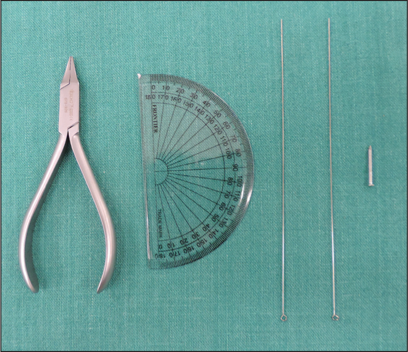 Figure 1
- Armamentarium used
The pin is attached to the center of the protractor after punching a hole through it. This can be simply done by heating the pin and passing it through and through at this point.
Two tiny helices of diameter slightly larger than that of the pin is formed at one end of both the round wires.
The two wires are then secured to the protractor by means of the pin by passing the helices over the pin.
The part of the pin that jets out through the other side is cut flush to the flat surface of the protractor. If the hole punched through the protractor using the pin is not larger than the diameter of the pin itself, then it would fit snugly and will not be displaced. However, in the case of a loose fit or if displacement of the pin is anticipated, then a small amount of self-cure acrylic can be added on the other side to secure it firmly. However, it is important to ensure that the acrylic does not come in contact with the wire helices as it will prevent free movement.
The ACAP is now ready to use and can be used repeatedly for any number of cephalometric analyses [Figure 2].
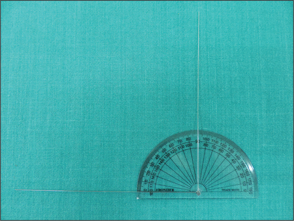 Figure 2
- Angular cephalometric analysing protractor
The device can be placed directly on the radiograph at suitable points for deriving the necessary angular measurements that are most commonly required by a clinician for diagnosis and treatment planning.
Illustrations of a few angles routinely used are presented to give a clear picture of how the ACAP can be used. For example: Gonial angle [Figure 3], saddle angle [Figure 4], SNA angle [Figure 5], SNB angle [Figure 6], upper incisor to SN angle [Figure 7], lower incisor to mandibular plane angle [Figure 8] and nasolabial angle [Figure 9]. Although the measurements may not be as precise as manual tracing, it certainly helps in quicker diagnosis and also due to its simplicity, should motivate more clinicians to derive cephalometric data before initiating treatment.
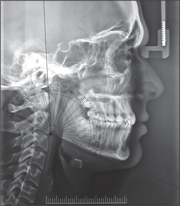
- Gonial angle
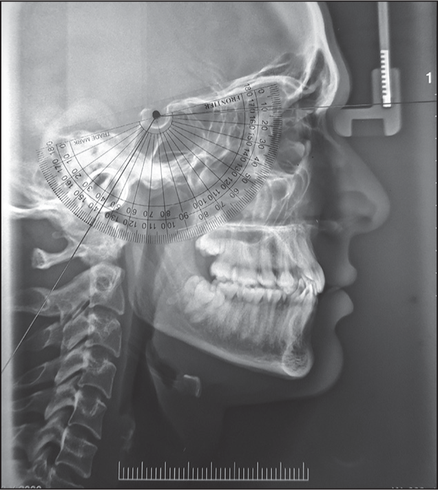
- Saddle angle
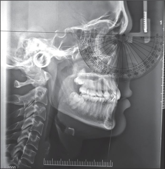
- SNA angle
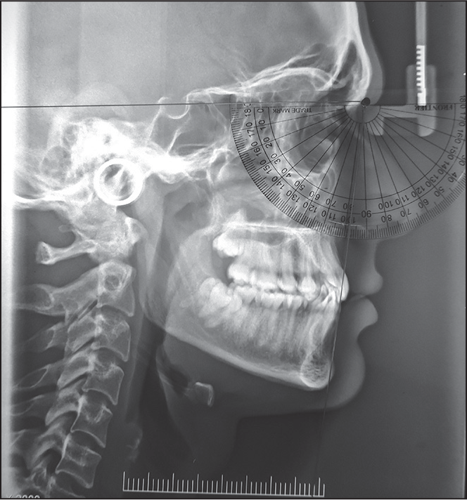
- SNB angle
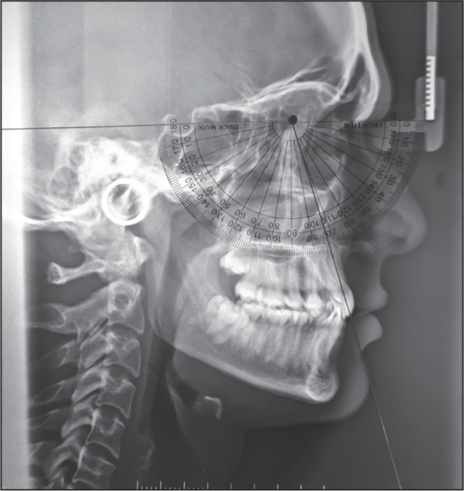
- Upper incisor to SN and SNB angle
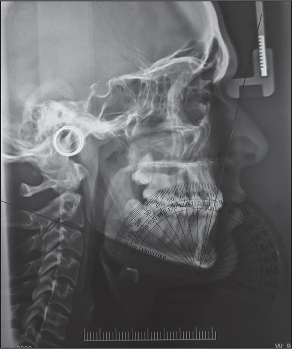
- Lower incisor to mandibular plane
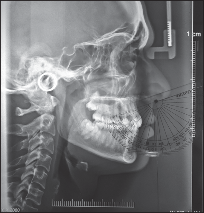
- Nasolabial angle
Source of Support:
Nil.
Conflict of Interest:
None declared.
References
- The reliability of head film measurements 2. Conventional angular and linear measures. Am J Orthod. 1971;60:505-17.
- [Google Scholar]
- Perspectives in the clinical application of cephalometrics. The first fifty years. Angle Orthod. 1981;51:115-50.
- [Google Scholar]
- Diagnosis and treatment planning for the surgical-orthodontic patient. Dent Clin North Am. 1990;34:361-84.
- [Google Scholar]
- The reliability and reproducibility of cephalometric measurements: A comparison of conventional and digital methods. Dentomaxillofac Radiol. 2012;41:11-7.
- [Google Scholar]


