

Translate this page into:
Accuracy of the infrazygomatic orthodontic bone screws digital planning and surgical guided positioning: A observational study


*Corresponding author: Paolo Manzo, Department of Orthodontics, University of Ferrara, Ferrara, Italy. paolo.manzo@gmail.com
-
Received: ,
Accepted: ,
How to cite this article: Manzo P, Paoletto E, Pellitteri F, Brucculeri L, Lombardo L. Accuracy of the infrazygomatic orthodontic bone screws digital planning and surgical guided positioning: An observational study. APOS Trends Orthod. 2024;14:85-90. doi: 10.25259/APOS_166_2023
Abstract
Objectives:
The objective of the study was to evaluate the accuracy of surgical guidance for the insertion of infrazygomatic (IFZ) miniscrews by means of a cone beam computed tomography (CBCT) evaluation.
Material and Methods:
Nine patients (five men and four women, from 14.8 to 41.4 years of age) were recruited for the study. Rhinoceros software was used to digitally plan the infrazygomatic miniscrew insertion with the INFRABSTEER method (INFRA-zygomatic and Buccal Shelf method), superimposing intraoral scans and a CBCT obtained at T0. No orthodontic movement was performed, and six months later a second CBCT was done (T1). Rhinoceros software was used to superimpose T0 and T1 files. Mesio-distal, bucco-palatal, and vertical linear discrepancy in cap and tip and an angular discrepancy between the mini-screws was measured. To check the repeatability of the measurements, the intraclass coefficient (ICC) was calculated. The significance level considered is P < 0.05. The ICCs are all >0.85 and significantly different from 0 (P < 0.05), indicating excellent repeatability of measurements.
Results:
The lowest mean linear distance was found to be the mesio-distal distance at the tip with 0.23 ± 0.241 mm. All linear distances produced an average distance of less than 0.5 mm, except for the bucco-palatal distance measured at the tip. The smallest angular measurement was obtained from the intersection of the axes of the miniscrews in the mesio-distal plane (1.58° ± 1.458°).
Conclusion:
The combination of a digitally planned and a template-guided surgical positioning procedure allows excellent control of infrazygomatic miniscrews insertion.
Keywords
Infrazigomatic crest
Infrazygomatic mini-screw
Surgical guide
Accuracy

INTRODUCTION
Anchorage control has always been one of the key aspects of orthodontic treatment planning and plays a key role in resolving the most complex cases. The use of temporary anchorage devices (TADs) is a tool available for the orthodontist. TAD has proven to be a useful addition in those treatments that require maximum anchorage control, limiting adverse effects that might occur with conventional anchorage.[1-7] Although they are widely used, nowadays miniscrew insertion is not a risk-free procedure, related to possible damage of roots or vascular-nervous structures, miniscrew bending or fracture, soft-tissue inflammation, and finally anchorage loss.[1,2,5]
Primary stability of the miniscrew and a good planning to avoid proximity to anatomical structures are the key factors to avoid possible complications and promote good biomechanics.[4,8]
The need to place miniscrews in the posterior area of the upper arch, to perform distalization movements,[9,10] intrusion of posterior dental areas[11], or space closure[5,7,12] is frequently arising in orthodontics.
However, the proximity to the roots can compromise the primary stability of the miniscrew, risking its loss or roots damage.
For this reason, a valid alternative could be the insertion of miniscrews in the infrazygomatic crest (IZC), a bridge of bone between the zygomatic process and the maxillary alveolar bone.[4,5,7] The safe zone indicated for the insertion of infrazygomatic (IFZ) miniscrews is generally located between the first and second upper molars in adults, and it’s bordered by a double-layered cortical bone, with the superior limit of the maxillary sinus floor.[1,4,13]
Sinus penetration is frequently reported during the insertion of miniscrews in the infrazygomatic area, although it is evidenced that a small perforation (<2 mm) can heal itself and create no undesirable side effects.[1,2]
Furthermore, it is agreed that a successful bicortical anchorage that is in the IZC, means to be very close to the sinus is crucial to achieve good primary stability of the miniscrews.[5]
Furthermore, for the wide intra- and inter-individual variety of the anatomy and of the thickness of the IZC, it can be critical to customize, by means of digital planning and Computer Aided Design/Computer Aided Manufacturing (CAD/CAM) guided surgery, the bone screw insertion in this bone area.[1]
The production of a guide based on a cone beam computed tomography (CBCT) of the patient allows a safe insertion of the miniscrew and to plan a safe insertion path, especially in those patients with complex anatomical situations. CBCTs enables precise positioning of the miniscrews, allowing for the planning of their placement in relation to both the maxillary sinus and adjacent tooth structures.[1,5,6,14]
The purpose of this study is to evaluate the accuracy of surgical guidance for the insertion of infrazygomatic miniscrews, comparing, by a digital superimposition, the planned IZC bone screws insertion and their clinically guided post-surgical position by means of a CBCT evaluation.
MATERIAL AND METHODS
After approval by the University of Ferrara Institutional Review Board, approval reference number - 07/2022, and informed release, nine patients consisting of five men and four women between the ages of 14.8 and 41.4 years were recruited for the study. Subjects selected for this study had to fulfill the following inclusion criteria: the planning of orthodontic treatment that required the use of one or two infra-zygomatic mini-screws and that the patient’s health condition allowed bimaxillary CBCT examination. Subjects with metal allergies, periodontal disease, or previous orthodontic treatment were excluded from the study.
For all patients, intraoral scans, intra- and extra-oral photographs for orthodontic treatment planning. Subsequently, a CBCT Green Vatech Tecnogaz (Green X Vatech, Korea; Multi-FOV sizes) was performed on all subjects in the same dental clinic to digitally plan infrazygomatic miniscrews insertion. For the digital planning, two different softwares were used, using the INFRABSTEER method (INFRA-zygomatic and Buccal Shelf method). This method consisted, firstly in the use of In vivo 6.0 and 3D Analysis software (Anatomage, San Jose, California) to process DICOM files from CBCTs and identify the optimal site direction, and depth of miniscrew insertion. In order to achieve a perfect fitting of the surgical guide on both teeth and soft tissue, the intraoral scan was superimposed on the CBCT files. The planning of the miniscrews was carried out by an orthodontic specialist with more than 20 years of experience, certified by the European Board of Orthodontics, together with a specialist technician with more than 10 years of experience.
Subsequently, Rhinoceros software (Robert McNeel and Associates, Seattle, WA) was used to virtually place the miniscrew between the first and second upper molars and to design a virtual surgical guide that will fit the full upper arch morphology for better stability.
Finally, the surgical guide was printed with the use of an Everes 3D printer (Sisma Piovene R. VI, Italy). The surgical guide presented a zygomatic extension slightly compressing the muco-gingival tissues of the IZC, in order to reduce the thickness and move away from the surgical pathway. The soft tissues and two cylindrical guides were placed in the buccal area to replicate the angle of insertion and prevent the screws from penetrating beyond the required depth [Figure 1]. The cylinder that is the screw-driving tool, had a window that allowed it to check the pick-up position and control the TAD insertion depth during the surgical procedure.
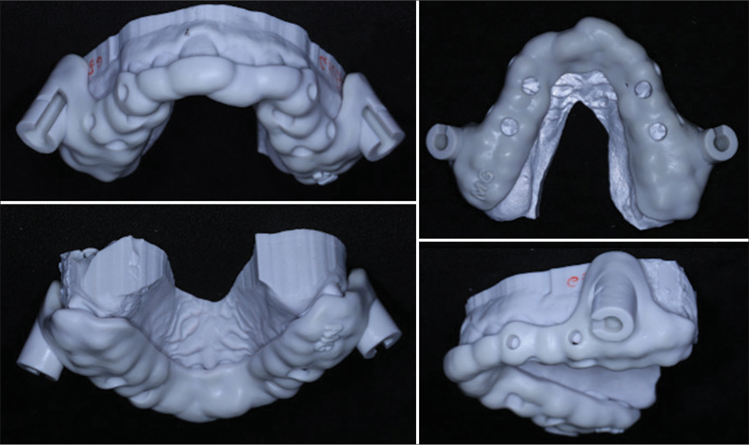
- The printed surgical guide for infrazygomatic miniscrews insertion.
Before the surgical procedure started, all patients were informed of the surgical risks and signed an informed consent.
First, the fitting and stability of the surgical guide was checked, and then following local anesthesia in the infrazygomatic area, 12 mm length and 2 mm diameter titanium miniscrews (Orthobone Screw, Taiwan) with a mushroom collar were inserted by means of a manual screw-driver.
After the surgical procedure performed by the same orthodontic specialist, and checking that a period of at least six months lasted from the T0 CBCT, a new CBCT was performed on all patients to avoid excessive radiation. During this period no orthodontic movement was performed.
CBCT files of all patients were collected, including the first digitally planned file with virtual miniscrew placement (T0) and the second file, after bone screw insertion (T1). Using the software Mimics (Version 20.0, Materialise©, Leuven, Belgium), DICOM files of the CBCTs were transformed into STL files.
To verify the accuracy of the surgical guide, Rhinoceros software was used so that the two STL files (T0 and T1) were first superimposed [Figure 2], and then measurements were performed. The models were first oriented on the X and Y axis, then the superimpositions were made on three reference points at the mesio-vestibular cusp of the upper first molar, inter-incisal point, and mesio-vestibular cusp of the upper first molar. In addition, the absence of tooth movement or appliance activation between T0 and T1 allowed a best-fitting superimposition.
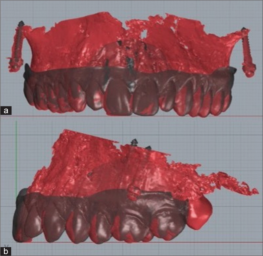
- (a) T0: Superimposition between programmed and (b) T1: real.
The vertical, mesio-distal, and bucco-palatal linear discrepancy in the miniscrews cap and in the tip, among the planned and the real miniscrews was then measured [Figure 3]. Finally, the vertical, mesio-distal, and buccopalatal angular discrepancy between the axis of the planned and the real miniscrews was measured. To limit a possible measurement error during point placement at the cap and tip, linear measurements were re-taken one week apart by the same operator.
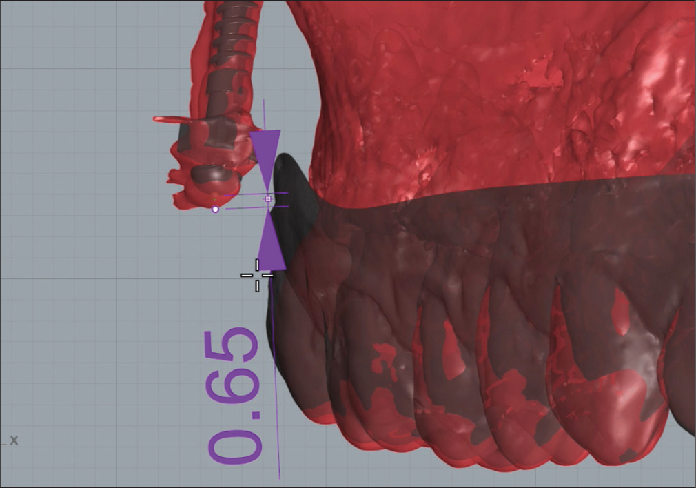
- Vertical linear measurement at the cap between T0 and T1. The purple arrows indicates the vertical millimeter distance between T0 and T1 in the cap.
Statistical analysis
Descriptive statistics of all variables were reported: being quantitative variables, mean, and standard deviation were calculated.
To check the repeatability of the measurements, the intraclass coefficient (ICC) was calculated from the two measurements taken.
For all tests, the significance level considered is P < 0.05. The analyses were conducted with IBM SPSS v28 software.
RESULTS
The final sample analyzed of the observational study consisted of nine patients (mean age 28.4 years) and 12 infrazygomatic miniscrews.
The descriptive analysis of the measurements made is given in [Table 1]. Since they are quantitative variables, the mean, standard deviation, median with 1° and 3° quartiles, and maximum and minimum values were studied.
| Variables | Mean±SD | Median (1°Q-3°Q) | Min–Max |
|---|---|---|---|
| Cap (mm) Vertical | 0.27±0.230 | 0.24 (0.05–0.46) | 0.02–0.65 |
| Cap (mm) Bucco-palatal | 0.33±0.264 | 0.37 (0.04–0.48) | 0.01–0.80 |
| Cap (mm) Mesio-distal | 0.41±0.368 | 0.30 (0.14–0.62) | 0.07–1.34 |
| Tip (mm) Vertical | 0.31±0.263 | 0.42 (0.17–0.64) | 0.00–0.79 |
| Tip (mm) Bucco-palatal | 0.54±0.481 | 0.47 (0.08–0.85) | 0.02–1.53 |
| Tip (mm) Mesio-distal | 0.23±0.241 | 0.14 (0.03–0.53) | 0.02–0.61 |
| Angular deviation (°) Vertical | 1.86±1.659 | 1.40 (0.83–2.58) | 0.20–5.80 |
| Angular deviation (°) Bucco-palatal | 3.43±3.869 | 2.00 (0.53–5.23) | 0.10–12.70 |
| Angular deviation (°) Mesio-distal | 1.58±1.458 | 1.25 (0.50–2.20) | 0.10–5.40 |
SD: Standard deviation.
The lowest mean linear distance was found to be the mesio-distal distance at the tip with 0.23 ± 0.241 mm, followed by the distance in the vertical direction at the cap with 0.27 ± 0.230 mm. All linear distances produced an average distance of <0.5 mm, except for the bucco-palatal distance measured at the tip (0.54 ± 0.481 mm).
As for the angular measurements, the smallest angle was obtained from the intersection of the axes of the miniscrews in the mesio-distal plane (1.58° ± 1.458°), while the largest is found in the bucco-palatal plane (3.43° ± 3.869°), reporting in fact a highest maximum value of 12.70°.
[Table 2] shows the intraclass correlation coefficient (ICC), which varies between 0 (no repeatability) and 1 (perfect repeatability). In this case, the ICCs are all >0.85 and significantly different from 0 (P < 0.05), indicating excellent repeatability of measurements.
| Variable | ICC |
|---|---|
| Cap | |
| Vertical | 0.996 |
| Bucco-palatal | 0.995 |
| Mesio-distal | 0.888 |
| Tip | |
| Vertical | 0.982 |
| Bucco-palatal | 0.999 |
| Mesio-distal | 0.993 |
ICC: Intraclass coefficient
DISCUSSION
Nowadays, the use of TADs for the resolution of complex orthodontic treatments requiring maximum anchorage is considered a routine procedure. However, the preservation of anatomical structures and primary stability of the miniscrew, are essential conditions to achieve maximum anchorage and safeguard the patient.[8]
To facilitate the safe insertion of TADs, a digital planning of the correct positioning of the miniscrew in all the three planes of the space can be carried out, whereby a surgical guide is designed and then created. In particular, when inserting miniscrew in the infrazygomatic area with a bicortical anchorage, care should be taken regarding the maxillary sinus, especially in adult patients, in whom the maxillary sinus is wider and therefore, sinus penetration for TADs is more likely.[3]
Through CBCT, sensitive structures can be identified the safe positioning of the infrazygomatic miniscrew can be designed and the high-precision surgical template individualized for each patient can be designed.[1,14]
CAD/CAM technology has been extensively utilized in the manufacturing of miniscrews surgical guides. (Su, Maino) The digitally designed 3D guide like the one described here can help the orthodontist to avoid any damage to anatomical structures, while reducing patient discomfort making more predictable the surgical bone screws positioning.[8] Indeed, the surgical guide design plays a key role in controlling the correct position of the miniscrew during its insertion: Two cylindrical guides are placed in the buccal area between the upper molars to replicate the angle of insertion and prevent the screws from penetrating beyond the planned depth.
In fact, the results obtained from this observational study showed that the linear and angular discrepancies in the vertical direction between the planned (T0) and the clinical actual position (T1) are extremely small: 0.27 ± 0.230 mm in the cap, and 0.31 ± 0.263 mm in the tip and 1.86° ± 1.659° of angle. Su et al.[1] investigated the accuracy of two different surgical guide designs for the insertion of infrazygomatic miniscrews, a template with a limiting ring to control verticality used in Group A, and a template with a semicircular structure to be able to change the angle of miniscrew insertion once the bony cortical layer has been engaged, for the Group B patients, and compared these results from the guided surgery with the results of a no-guided surgery in the Group C patients. The results obtained from this observational study showed that in Group A, there was a greater vertical control with an average discrepancy of 0.62± 0.67 mm in the cap, 0.55 ± 0.61 mm in the tip, and 9.52 ± 10.42 mm in angulation. On the other hand, Group B produced more than 1 mm of linear vertical discrepancy at the cap and just under 1 mm at the tip, with an angulation discrepancy in the vertical direction of more than 10° on average. Moreover, Group C of patients with no-guided surgery proved to be the group with higher values of both cap and tip standard deviation and angular deviations, reaching 3.96 mm ± 5.81mm, 3.29 mm ± 2.43 mm, and 14.69° ± 8.21°, respectively. These results demonstrate how the insertion of infrazygomatic miniscrews without the aid of a surgical guide can imply an imprecise positioning and can be dangerous for the surrounding anatomical structures.
In this regard, considering the studies by Kravitz and Kusnoto[2] and Jia et al.[5] where the possible complications of miniscrews insertion are studied, both supported the importance of vertical control to avoid excessive sinus perforation. In particular, Jia, who studied CBCTs of patients before and after infrazygomatic miniscrew insertion, pointed out how the overall success of miniscrews in this region was 96.7%, but 78.3% penetrated into the sinus, and when the penetration was >1 mm, the thickening of Schneider’s membrane was 88.2%.
Mesio-distal and bucco-palatal control during the insertion of infrazygomatic miniscrews allowed the preservation of possible adjacent tooth structures, especially the mesio-buccal root of the upper second molar.[13] The results obtained showed that through the design of this surgical guide, an excellent control of the mesio-distal and bucco-palatal linear distance is achieved, with a linear discrepancy <0.5 mm, except for the bucco-palatal distance at the tip where an average distance of 0.54 ± 0.481 mm is reported and the angulation in the same direction with 3.43° ± 3.869°. The results are in line with those obtained in the study by Bae et al.[6] who reported a mesio-distal deviation in the cap of 0.29 mm and 0.21 mm in the tip. Mesio-distal and bucco-palatal control of the miniscrews during insertion was achieved by a cylindrical system with only a small window to verify the position of the pick-up driver during insertion. Moreover, Su et al.,[1] demonstrated how a semicircular structure that does not cover the entire circumference of the pick-up can allow for the arising of angle changes during the insertion of the miniscrew. The study[1] provides discrepancies in Group B patients in the inclination of the miniscrew, ranging in the mesio-distal direction from 2.38 ± 1.66 mm of linear discrepancy in the cap, to a 3.32 ± 1.45 mm in the tip. Furthermore, as for deviations in cap and in the tip for vertical movements, for mesio-distal and bucco-palatal movements the C group with no-guided surgery produced greater deviations than the groups using a surgical guide. Indeed, the insertion of the miniscrews without the use of a surgical guide produced deviations ranging from a minimum value of 2.77 mm ± 2.09 mm in the mesio-distal direction in the cap, up to 3.86 mm ± 5.27 mm also in the cap in the bucco-palatal direction.
It must also be considered that there are factors that may contribute to errors in the placement of IZC miniscrews, such as possible inaccuracies in the printing of the surgical guide. However, Ma et al.[15] in their study reported that deviation of measurements was considered acceptable in making a surgical guide from a CBCT file. However, Pozzan et al.[16] reported that laboratory procedures cannot provide a significant role in the degree of deviations of miniscrew placement. Conversely, the clinical steps have a bigger influence and need to be carefully evaluated, for these reasons more clinical studies on the accuracy of miniscrews placement must be carried out.
Despite the satisfactory results obtained in this observational study, the low number of patients included must be taken into consideration, this could influence the results obtained. For this reason, further studies are needed to increase the study sample and confirm the results obtained.
CONCLUSION
The combination of a digitally planned and a template-guided surgical positioning procedure with the INFRABSTEER method, allows excellent control of infrazygomatic miniscrews insertion in the mesio-distal and bucco-palatal directions and especially in the vertical direction. Here there is an average discrepancy at the tip of 0.31 mm between the planned and real bone screw position, ensuring excellent positioning of the miniscrew and preservation of the surrounding anatomical structures.
Ethical approval
The study is approved by the Institutional Review Board at University of Ferrara number 07/2022, dated 23rd June 2022.
Declaration of patient consent
The authors certify that they have obtained all appropriate patient consent.
Conflicts of interest
Dr. Luca Lombardo is on the editorial board of the Journal.
Use of artificial intelligence (AI)-assisted technology for manuscript preparation
The authors confirm that there was no use of artificial intelligence (AI)-assisted technology for assisting in the writing or editing of the manuscript and no images were manipulated using AI.
Financial support and sponsorship
Nil.
References
- Accuracy of two orthodontic mini-implant templates in the infrazygomatic crest zone: A prospective cohort study. BMC Oral Health. 2022;22:252.
- [CrossRef] [PubMed] [Google Scholar]
- Risks and complications of orthodontic miniscrews. Am J Orthod Dentofacial Orthop. 2007;131:S43-51.
- [CrossRef] [PubMed] [Google Scholar]
- Success of infrazygomatic crest bone screws: Patient age, insertion angle, sinus penetration, and terminal insertion torque. Am J Orthod Dentofacial Orthop. 2022;161:783-90.
- [CrossRef] [PubMed] [Google Scholar]
- Miniscrew insertion sites of infrazygomatic crest and mandibular buccal shelf in different vertical craniofacial patterns: A cone-beam computed tomography study. Korean J Orthod. 2021;51:387-96.
- [CrossRef] [PubMed] [Google Scholar]
- Influence of orthodontic mini-implant penetration of the maxillary sinus in the infrazygomatic crest region. Am J Orthod Dentofacial Orthop. 2018;153:656-61.
- [CrossRef] [PubMed] [Google Scholar]
- Accuracy of miniscrew surgical guides assessed from cone-beam computed tomography and digital models. Am J Orthod Dentofacial Orthop. 2013;143:893-901.
- [CrossRef] [PubMed] [Google Scholar]
- A computed tomographic image study on the thickness of the infrazygomatic crest of the maxilla and its clinical implications for miniscrew insertion. Am J Orthod Dentofacial Orthop. 2007;131:352-6.
- [CrossRef] [PubMed] [Google Scholar]
- A three-dimensional digital insertion guide for palatal miniscrew placement. J Clin Orthod. 2016;50:12-22.
- [Google Scholar]
- Three-dimensional evaluation on the effect of maxillary dentition distalization with miniscrews implanted in the infrazygomatic crest. Implant Dent. 2018;27:22-7.
- [CrossRef] [PubMed] [Google Scholar]
- Safe zones for miniscrews in maxillary dentition distalization assessed with cone-beam computed tomography. Am J Orthod Dentofacial Orthop. 2017;151:500-6.
- [CrossRef] [PubMed] [Google Scholar]
- Skeletal anchorage system for open-bite correction. Am J Orthod Dentofacial Orthop. 1999;115:166-74.
- [CrossRef] [PubMed] [Google Scholar]
- Effectiveness of orthodontic miniscrew implants in anchorage reinforcement during en-masse retraction: A systematic review and meta-analysis. Am J Orthod Dentofacial Orthop. 2017;151:440-55.
- [CrossRef] [PubMed] [Google Scholar]
- Safe sites for orthodontic miniscrew insertion in the infrazygomatic crest area in different facial types: A tomographic study. Am J Orthod Dentofacial Orthop. 2022;161:37-45.
- [CrossRef] [PubMed] [Google Scholar]
- Surgical positioning of orthodontic mini-implants with guides fabricated on models replicated with cone-beam computed tomography. Am J Orthod Dentofacial Orthop. 2007;131:S82-9.
- [CrossRef] [PubMed] [Google Scholar]
- The accuracy of a 3D printing surgical guide determined by CBCT and model analysis. J Adv Prosthodont. 2018;10:279-85.
- [CrossRef] [PubMed] [Google Scholar]
- Accuracy of the digital workflow for guided insertion of orthodontic palatal TADs: A step-by-step 3D analysis. Prog Orthod. 2022;23:27.
- [CrossRef] [PubMed] [Google Scholar]


