

Translate this page into:
An edge over diagnostic setup
Address for correspondence: Dr. Ashish Kumar Singh, Department of Orthodontics and Dentofacial Orthopedics, Manav Rachna Dental College, Faridabad, Haryana, India. E-mail: drashish.singh@gmail.com
This article was originally published by Wolters Kluwer and was migrated to Scientific Scholar after the change of Publisher.
Abstract
Diagnostic setup proposed by H.D. Kingsley serves as a practical aid in treatment planning and diagnosis. These setups have some inherent shortcomings. A simple technique of duplication of the setups in dental stone can solve problems encountered before as well as provide many other advantages over the conventional procedure. The diagnostic setup is prepared by the conventional method [Figure 1]. An alginate impression is then taken of the setups and poured in dental stone to obtain the derived treatment model [Figure 2]. The same setup can now be further modified for alternate lines of treatment. Subsequently models could then be obtained as required [Figure 3].
Keywords
Diagnostic setup
study models
kesling

Introduction
Diagnostic setup proposed by Kesling[1] serves as a practical aid in treatment planning and diagnosis. Some of the advantages of the conventional setup[2] include:
To determine and visualize the resultant occlusion
Tooth size – arch length discrepancies can be better optimized
Useful aid in asymmetric extraction and combined surgical orthodontic treatment planning.
These setups have some inherent shortcomings.[3,4] First, patients for whom multiple treatment options are thought of, multiple diagnostic setups need to be made which would be very laborious and time-consuming. Furthermore, the patients may not fully appreciate the wax setups shown to them.
A simple technique of duplication of the setups in dental stone can solve problems encountered before as well as provide many other advantages over the conventional procedure.
Procedure
The diagnostic setup is prepared by the conventional method [Figure 1]. An alginate impression is then taken of the setups and poured in dental stone to obtain the derived treatment model [Figure 2]. The same setup can now be further modified for alternate lines of treatment.[5] Subsequently, models could then be obtained as required [Figure 3].

- Pretreatment study models and diagnostic setup
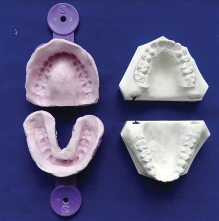
- Alginate impression of Kesling setup and derived treatment model
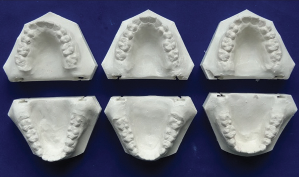
- Multiple treatment models of proposed treatment plans
The advantages of this simple technique are:
Better visualization of treatment results for the operator as well as the patient
Multiple treatment models for the same patient
More accurate fabrication of customized archwires
Plaster models are dimensionally more stable as compared to the wax setups
Greater patient motivation
More precise bracket positioning of lingual appliances
Accurate fabrication of tooth positioners if required.
Financial support and sponsorship
Nil.
Conflicts of interest
There are no conflicts of interest.
References
- The philosophy of the tooth positioning appliance. Am J Orthod Oral Surg. 1945;31:297-304.
- [CrossRef] [Google Scholar]
- Easy wax setup technique for orthodontic diagnosis. J Clin Orthod. 2000;34:140-4.
- [CrossRef] [Google Scholar]


