Translate this page into:
Evaluation of the Effects of the Chincup Appliance on the Craniofacial Structures by the Finite Element Analysis
Address for correspondence: Dr. Beril Demir Karamanli, Istanbul University, Faculty of Dentistry, Department of Orthodontics, 34093, Capa-Fatih, Istanbul, Turkey. E-mail: berilkaramanli@gmail.com
This article was originally published by Wolters Kluwer and was migrated to Scientific Scholar after the change of Publisher.
Abstract
Aims
The aim of this study is to evaluate the effects of the chincup appliance used in the treatment of Class III malocclusions, not only on the mandible or temporomandibular joint (TMJ) but also on all the craniofacial structures.
Materials and Methods
Chincup simulation was performed on a three-dimensional finite element (FE) model. 1000 g (500 g per side) force was applied in the direction of chin-condyle head. Nonlinear FE analysis was used as the numerical analysis method.
Results
By the application of chincup, stresses were distributed not only on TMJ or mandible but also on the circummaxillary sutures and other craniofacial structures.
Conclusions
Clinical changes obtained by chincup treatment in Class III malocclusions are not limited by only mandible. It was seen that also further structures were affected.
Keywords
Chincup
craniofacial system
finite element analysis
Introduction
There are two different treatment approaches in the treatment of Class III malocclusions. One of them is early orthopedic treatment before the growth peak, and the other one is orthognathic surgery or camouflage treatment after the growth period. Treatment type must be chosen due to the affected skeletal component in skeletal Class III malocclusions.[1]
Chincup appliance takes place in the treatment of Class III malocclusions due to prognathic mandible for many years. Some researches, evaluating the effects of the chincup appliance, reported that mandibular growth was inhibited[2-5] or mandibular shape was changed[2] by chincup therapy; however, many researches stated that mandibular growth was not affected by the retraction force applied on the mandible by chincup.[6-10] It is generally accepted that, by the chincup therapy, mandibular growth is not inhibited, but mandibular growth direction is changed to downward and backward.[7-10]
Several studies have indicated that the chincup not only has effects on the growth of the mandible but also on cranial base structures as well.[4,5,11,12] Ritucci and Nanda[11] reported that the chincup causes a closure of the cranial flexure angle (N-S-Ba) associated with the inhibited posterior growth of the posterior cranial base at basion and the upward movement of sella. This positional change of the temporomandibular joint (TMJ) may affect the position of the mandible directly. Wendl et al.[4] reported that in long term, follow-up of early chincup treatment effected mandibular body length, effective mandibular length, SNB, ANB, and gonial angle and this type of early treatment did not have adverse impact on the TMJs.
Although studies have evaluated the biomechanical effects of various chincup force vectors on the mandible and TMJ,[13-15] no data are available about the biomechanical effects of the appliance on whole craniofacial system. Therefore, our aim in this study was to evaluate the biomechanical effects of chincup treatment on the craniofacial structures using a 3-dimensional (3D) finite element (FE) model.
Materials and Methods
Chincup simulation was performed using a 3D FE model. 3D model of the craniofacial complex which was obtained by 3D optic scanning of the craniofacial bones were provided from a company (21st Century Solutions Ltd., Suite 31, Don House, 30-38 Main Street, Gibraltar). Size of the 3D model was equivalent of the size of an adolescent skull with Class III malocclusion. 3D model was saved as digital imaging and communications in medicine file and then imported to CATIA V5 R14 (Dassault Systemes) software for 3D reconstruction. TMJ discs and circummaxillary sutures were designed manually using CATIA software. The reconstructed geometry of craniomaxillary complex was exported in stereolithography (STL) file format. The STL file was imported into SAMCEF Field (SAMTECH), which was used to generate a volume mesh from the 3D geometry of the craniofacial complex. The model was meshed using 539262 tetrahedral elements and 135823 nodes. The mechanical properties of the bones, teeth, sutures, and TMJ discs were defined according to experimental data from previous studies [Table 1].[16,17] The materials in the analysis were assumed to be nonlinear and TMJ discs and sutures viscoelastic. Kelvin model was used as the viscoelastic material model.[18]
| Young modulus (MPa) | Poisson ratio (v) | |
|---|---|---|
| Cortical bone | 13,700 | 0.3 |
| Teeth | 20,290 | 0.3 |
| Cartilage layers | 0.79 | 0.49 |
In the simulation of chincup application, a 1000 g (500 g per side) force was directed from the chin to the condyle head. Analysis was performed for 300 s.
Results
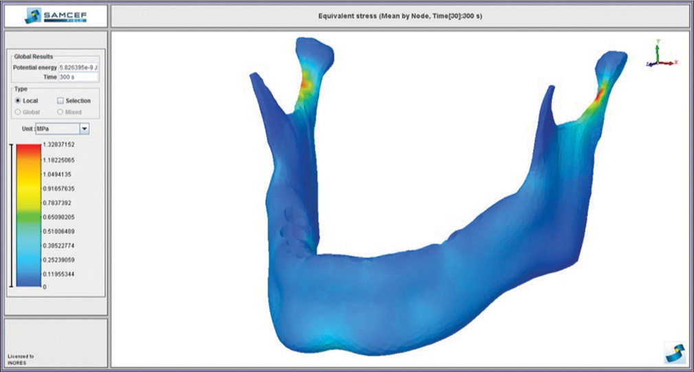
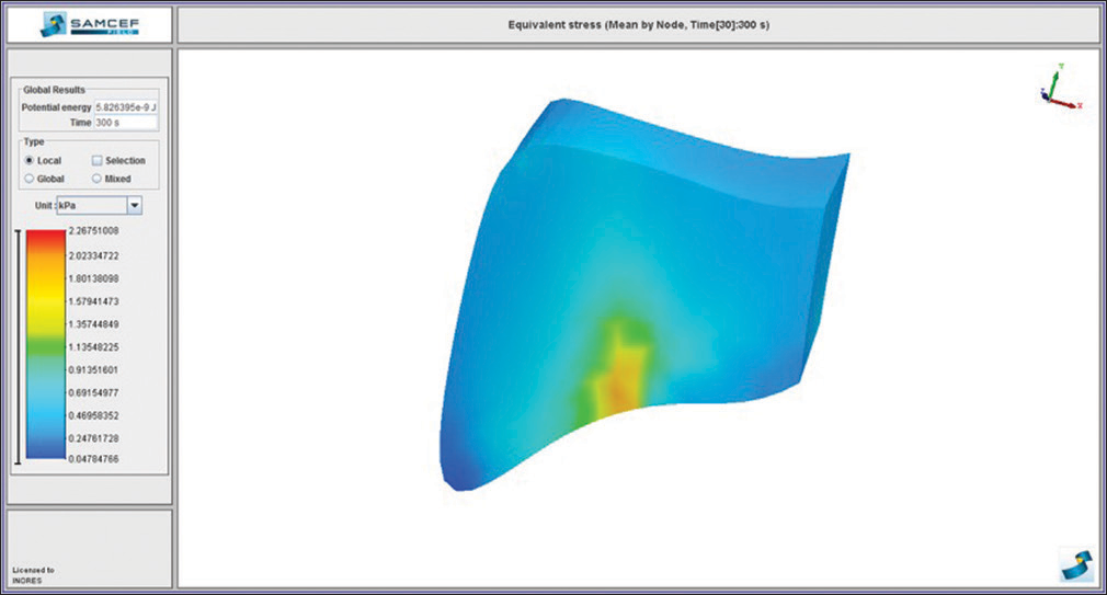
In chincup treatment simulation, von Mises stresses were seen in zygomaticomaxillary suture (2.42 MPa), frontomaxillary suture (1.80 MPa), condyle necks (1.32 MPa), nasomaxillary suture (0.72 MPa), zygomaticotemporal suture (0.68 MPa), articular discs (0.16 MPa), pterygopalatine suture (0.14 MPa), temporal bone (0.07 MPa), and frontal process of the maxilla (0.0037 MPa) in descending order [Figures 1-10].

- Von Mises stress distribution on the craniofacial system

- Von Mises stress distribution on the mandible

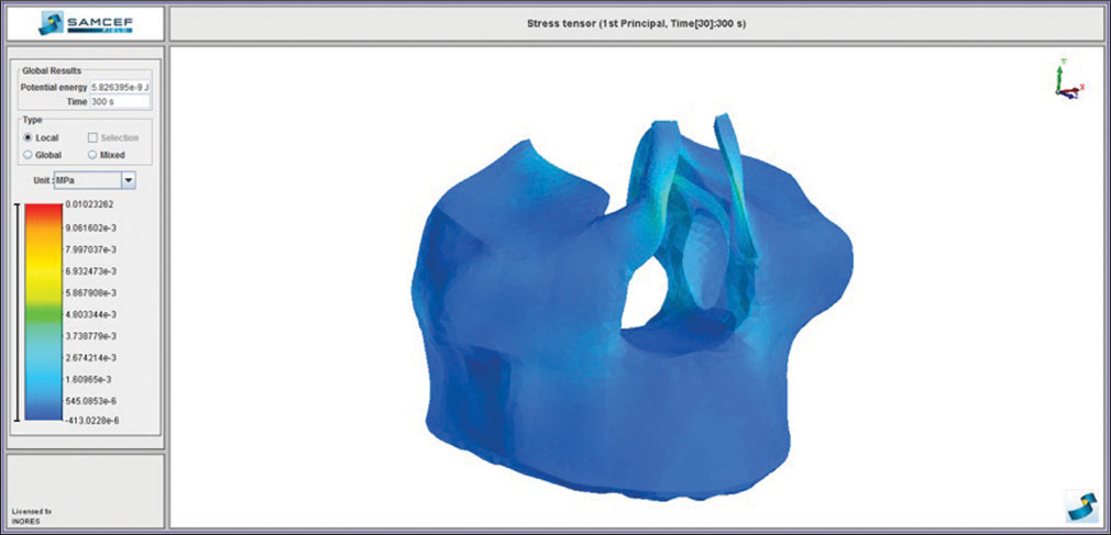
- Von Mises stress distribution on the maxilla

- Von Mises stress distribution on the temporal bones

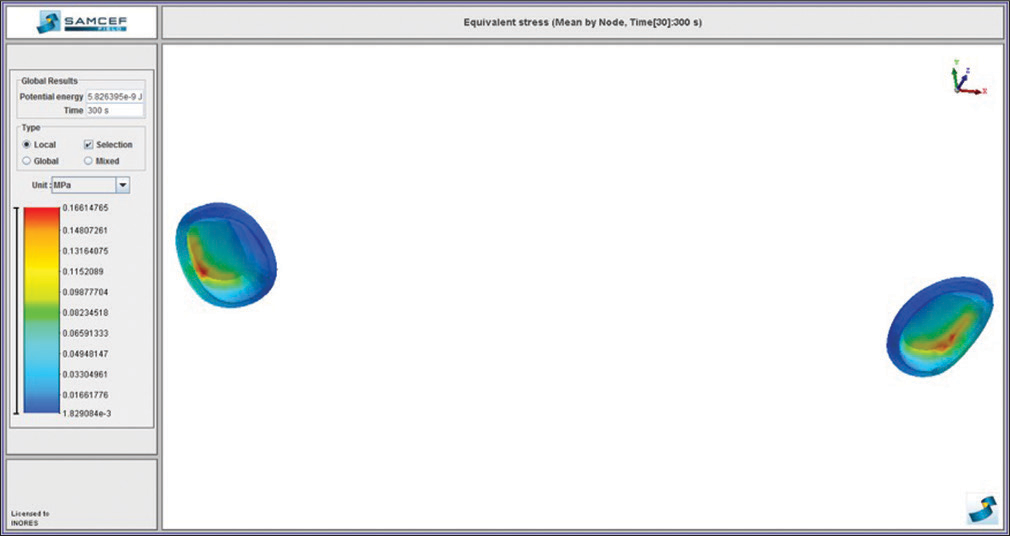
- Von Mises stress distribution on the temporomandibular joint discs

- Von Mises stress distribution on the zygomaticotemporal suture

- Von Mises stress distribution on the zygomaticomaxillary suture

- Von Mises stress distribution on the pterygopalatine suture

- Von Mises stress distribution on the frontomaxillary suture

- Von Mises stress distribution on the nasomaxillary suture
Uniform von Mises stress distributions were observed in the maxilla. Stresses were higher in frontal process of the maxillary bone [Figure 3].
By the force applied to the chin, stresses were distributed in the posterior edge of the glenoid fossa and the zygomatic edge of the temporal bone [Figure 4].
Von Mises stresses in the TMJ discs were higher in the upper posterolateral sides of the discs [Figure 5].
Higher stresses in the mandible were observed in the condyle necks [Figure 2].
In circummaxillary sutures, higher stresses were observed in zygomaticomaxillary suture, frontomaxillary suture, nasomaxillary suture, zygomaticotemporal suture, and pterygopalatine suture in descending order [Figures 6-10].
Discussion
By the 1000 g force applied to the chin, high von Mises stresses in the mandible were observed on the condyle necks [Figure 2]. Stresses on the condyle necks appear to be similar to the flexion effect which occurs when a force is applied on the long bones horizontally. In the mandible, similar flexion occurs with the condyle necks as the flexion center. This flexion effect is thought to be effective on the remodeling of the mandible.[13,15,19]
Basciftci et al.[15] applied a chincup force with 500 g magnitude in three different directions as chin-condyle head, chin-coronoid process, and chin in front of coronoid process and evaluated the stress distribution on the mandible. The highest von Mises stresses were seen on the condyle region and posterior of the ramus, and stresses tended to rise when the force vector moved away from condyle head. Average von Mises stresses on the condyle head was 0.069 MPa when the force passed through the condyle head and tended to reduce through the coronoid process. In our study, force vector passed through the condyle head and highest stresses were observed on the condyle neck (1.32 MPa).
Tanne et al.,[13] in their FE study, applied a 400 g force in the direction of chin-condyle head. Tensile stresses were observed on the outer surface of the mandible, and compressive stresses were observed inside of the mandible. Researchers reported that this stress difference on the mandible was responsible of the morphologic changes on the bone by the chincup therapy. Stresses on the corpus of the mandible were higher than the stresses seen on the condyle head.
In our study, by the force applied to the chin, almost uniform stress distribution on the maxilla was observed [Figure 3]. Mildly higher von Mises stresses were observed on the frontal process of the maxilla. These findings showed us that chincup force was transmitted to the further structures by the glenoid fossa and zygomatic bone. In the literature, it is generally supported that chincup was not effective on the maxilla;[20,21] however, in some studies, it is reported that by the chincup therapy mild rising on the maxillary, vertical sizes and cranial base occur.[11,22,23]
Ritucci and Nanda[11] applied 500 g force in the direction of chin-condyle neck and compared the effects of the chincup therapy with the control group. The results of this study indicate that chincup causes a closure in the N-S-Ba angle, inhibition of the posterior growth of the Ba point, and imposes a vertical growth tendency on the points nasion and sella. The chincup significantly inhibits anterior and posterior vertical maxillary growth and growth of the upper anterior facial height. Because the development of vertical posterior facial height is inhibited more than anterior facial height, a clockwise rotation of the maxilla occurs. In our study, distribution of the von Mises stresses, especially on the frontal process of the maxilla, agrees with the studies that support that chincup was effective on the maxilla.
In chincup simulation, von Mises stresses on the temporal bone were higher on the glenoid fossa and zygomatic edge of the bone [Figure 4]. This finding supports that the force applied to the chin is transmitted to the glenoid fossa and temporal bone through articular discs.
As the force applied to the chin is transmitted to the condyle head, condyle head pressures on the TMJ disc and a deformation occurs. This deformation caused the von Mises stresses to be higher on the posterolateral sides and the convexity of the discs [Figure 5]. In Tanne et al.’s[14] study, 400 g force was applied to the chin through the condyle head in 50° angle. Condyle heads pressured on the posterior of the discs and caused a tensile stress on the anterior of the discs. The reason for this is that the force applied was in posterior direction and caused the condyle heads move backward and created a compressive stress on the posterior of the discs. In our study, condyle heads created compression on the convexity of the discs which were in the direction of the force vector.
Conclusions
In our study, effects of the chincup appliance on the craniofacial structures were evaluated using the FE analysis. Contrary to the expectations, chincup was found effective not only on the mandible but also on the further craniofacial structures.
Financial support and sponsorship
This study was financially supported by TÜBİTAK (The scientific and technological research council of Turkey) Project number: 13770.
Conflicts of interest
There are no conflicts of interest.
References
- Cephalometric variables predicting the long-term success or failure of combined rapid maxillary expansion and facial mask therapy. Am J Orthod Dentofacial Orthop. 2004;126:16-22.
- [Google Scholar]
- Chincup treatment modifies the mandibular shape in children with prognathism. Am J Orthod Dentofacial Orthop. 2011;140:38-43.
- [Google Scholar]
- A roentgenocephalometric study of skeletal changes during and after chincup treatment. Am J Orthod. 1984;85:341-50.
- [CrossRef] [Google Scholar]
- Long-term skeletal and dental effects of facemask versus chincup treatment in class III patients: A retrospective study. J Orofac Orthop. 2017;78:293-99.
- [CrossRef] [PubMed] [Google Scholar]
- Comparisons of two protocols for the early treatment of class III dentoskeletal disharmony. Eur J Orthod. 2016;38:51-56.
- [Google Scholar]
- Efficacy of short-term chincup therapy for mandibular growth retardation in class III malocclusion. Angle Orthod. 2011;81:162-68.
- [CrossRef] [PubMed] [Google Scholar]
- Facial growth of skeletal class III malocclusion and the effects, limitations, and long-term dentofacial adaptations to chincap therapy. Semin Orthod. 1997;3:244-54.
- [Google Scholar]
- Early treatment of class III incisor relationship using the chincap appliance. Eur J Orthod. 1993;15:371-6.
- [CrossRef] [Google Scholar]
- Chin cap force to a growing mandible. Long-term clinical reports. Angle Orthod. 1984;54:93-122.
- [Google Scholar]
- Long-term effect of the chincap on hard and soft tissues. Eur J Orthod. 1999;21:291-8.
- [CrossRef] [PubMed] [Google Scholar]
- The effect of chincup therapy on the growth and development of the cranial base and midface. Am J Orthod Dentofacial Orthop. 1986;90:475-83.
- [Google Scholar]
- Retrospective 25-year follow-up of treatment outcomes in angle class III patients: Early versus late treatment. J Orofac Orthop. 2017;78:201-10.
- [CrossRef] [PubMed] [Google Scholar]
- Biomechanical changes of the mandible from orthopaedic chincup force studied in a three-dimensional finite element model. Eur J Orthod. 1993;15:527-33.
- [Google Scholar]
- Stress distribution in the temporomandibular joint produced by orthopedic chincup forces applied in varying directions: A three-dimensional analytic approach with the finite element method. Am J Orthod Dentofacial Orthop. 1996;110:502-7.
- [Google Scholar]
- Biomechanical evaluation of chincup treatment with various force vectors. Am J Orthod Dentofacial Orthop. 2008;134:773-81.
- [Google Scholar]
- Stress distribution in the temporomandibular joint after mandibular protraction: A 3-dimensional finite element method study. Part 1. Am J Orthod Dentofacial Orthop. 2009;135:737-48.
- [Google Scholar]
- Association between mechanical stress and bone remodeling. J Osaka Univ Dent Sch. 1990;30:64-71.
- [Google Scholar]
- Three-dimensional finite-element model of the human temporomandibular joint disc during prolonged clenching. Eur J Oral Sci. 2006;114:441-8.
- [Google Scholar]
- Static vs. dynamic loads as an influence on bone remodelling. J Biomech. 1984;17:897-905.
- [CrossRef] [Google Scholar]
- Treatment effects of the light-force chincup. Am J Orthod Dentofacial Orthop. 2010;138:468-76.
- [CrossRef] [PubMed] [Google Scholar]
- Dentofacial changes in patients with class III malocclusions treated by a combination of activator and chin-cup appliances. Aust Orthod J. 1997;14:225-8.
- [Google Scholar]
- Long-term evaluation after chincap treatment. Eur J Orthod. 1995;17:135-41.
- [CrossRef] [PubMed] [Google Scholar]
- Stability of changes associated with chincup treatment. Angle Orthod. 1996;66:139-45.
- [Google Scholar]


