Translate this page into:
Instant Virtual Treatment Outcome Using PowerPoint
Address for correspondence: Dr. Kavitha S. Iyer, Department of Orthodontics and Dentofacial Orthopedics, Ragas Dental College and Hospital, Chennai, Tamil Nadu, India. E-mail: drkavithaiyer@gmail.com
This article was originally published by Wolters Kluwer and was migrated to Scientific Scholar after the change of Publisher.
Abstract
The aim of this article is to introduce a faster and even simpler method where the patient can readily visualize the treatment outcome than the currently available methods. This article demonstrates how PowerPoint software can be used to generate instant virtual treatment outcomes (IVTOs) at the chairside using the patient’s digital photographs. The generated two-dimensional skeletal and dental IVTO provides the necessary communication aid in patient motivation and education. Furthermore, it serves as making a tremendous leap forward, toward informed consent.
Keywords
Dental virtual outcome
patient counseling
surgical outcome
treatment outcome
virtual treatment outcome
Introduction
Orthodontic imaging software is designed for use by specialized dental practices for capturing, storing, and presenting patient images and assisting in treatment planning and case diagnosis. The speed of computerized cephalometric programs has helped streamline the laborious manual measurement of patient cephalograms and has also hastened the creation of the visualization treatment objective.[1] Informed consent has taken a tremendous leap forward with the screen view of the end result provided by treatment planning software to the patient that is far more aptly illustrative than the old wax setup. No longer does the final result exist only in the mind of the orthodontist, or as a comparison with samples of previously treated cases.[2]
Imaging software effortlessly manages patient pictures and X-ray images with a set of powerful and user-friendly tools for conveying treatment plans. They help easily communicate ideas through a more colloquial and less technical language with greater patient realization and acceptance for treatment. Treatment visualization approach is classified as image editing systems and photo-cephalometric treatment visualization systems, and it can be depicted in two-dimension (2D) or three-dimension (3D).[3]
FDA-Cleared Class II medical devices such as Dolphin imagining software (Dolphin Imaging and Management Solutions, Eton Ave., Chatsworth, CA, USA) have been used for over a decade by many practitioners as an effective tool for aiding in virtual treatment and to measure the outcomes. This software can provide possible treatment outcome, simulate surgical results, and allow the operator, completely refine predicted treatment results. The soft-tissue outline can be easily detailed in the image with a drawing tool (blend brush) for image smoothness. Recently, SureSmile (OraMetrix, Inc., Hillcrest, Dallas, TX, USA) technology has allowed the practitioner to perform accurate, 3D treatment simulations based on data supplied by its handheld OraScanner (OraMetrix, Inc., Hillcrest, Dallas, TX, USA). This state-of-the-art technology has taken patient education, treatment planning, and appliance to newer dimension.[4,5] However, the results produced by these software’s diagnostic and treatment planning tools are dependent on the interpretation of trained and experienced practitioners.[6] A cheaper, faster, and even simpler method where the patient can readily visualize the treatment outcome was needed.
Use of PowerPoint (Office 2016, Microsoft Corporation, Redmond, WA, USA) to manage digital photographs and case presentations and create customized documents for the orthodontic office has been well reported in the literature.[7-12] It has a myriad of features that can enhance the delivery of content with proper meaning and enable the user to customize their presentations. Its ability to incorporate multimedia files and portability of records has become the de facto presentation software for the Information Age.[13] This article presents a fast method to develop a 2D dental and skeletal virtual treatment outcome using patient photographs with PowerPoint during the patient’s consultation visit.
Procedure
Any computer operating system installed with Microsoft Office PowerPoint 2007 or higher version software is required for rendering instant virtual treatment outcome (IVTO). Digital images stored within the operating system or from an external device/cloud storage can be imported into the operating system for generating IVTO.
The image to be converted to IVTO is imported into the PowerPoint software using insert option in the view tab on the toolbar or by simple drag and drop option from the file folder into a PowerPoint slide [Figure 1]
 Figure 1
- Image imported into Powerpoint
Additional geometric references such as the ruler, gridlines, and guides can be added from the view tab in the toolbar into the slide to help with image orientation
Use diagonal stretching to increase or decrease the size of the picture without changing the image magnification
Make copies of the image by clicking CTRL-C and CTRL-V (for Windows operation system) or Command + C to copy, then Command + V to paste (for Mac OS) in the same slide. Use the image copies to crop, stretch, rotate, or flip it horizontally from the format tab in the view option of the toolbar as per the superimposition requirement over the original image to make the desired virtual treatment outcome
Once the image is finalized, select all the images to be merged into a single IVTO image
-
Generated dental and skeletal IVTO images are compared with the pretreatment image for easy communication with the patient for treatment outcome visualization and acceptance.
Generating the Instant Virtual Treatment Outcomes
Case 1
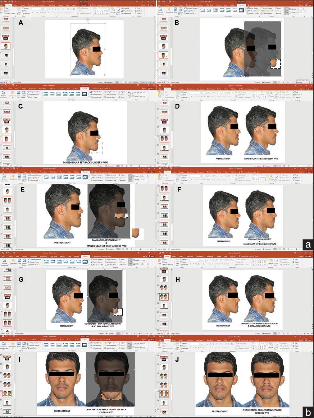
Surgical IVTO was done for a 24-year-old male patient with skeletal Class III profile. Three treatment plans were incorporated in the skeletal IVTO. Extraoral digital photograph of the patient was imported at chairside into the PowerPoint 2016 software. First, for the mandibular setback and maxillary advancement, two copies of the extraoral profile photograph were cropped, one in the lower two-third face at the level of lip commissure extending below for mandibular setback, and in another photograph, the cropping for upper one-third of lower face was done at lip commissure extending above to the subnasale for maxillary advancement. The cropped pictures were superimposed based on profile improvement. In the second treatment plan for only the mandibular setback, the previous setback cropping was superimposed on the mandibular segment without maxillary advancement.
In the third treatment plan of reduction and setback genioplasty, only the chin part of the photograph was cropped and superimposed superiorly and posteriorly. The chairside generated IVTO helped the patient to go with the first treatment plan of maxillary advancement and mandibular setback surgery [Figure 2]. The patient was informed that a further evaluation was needed to finalize the treatment objectives and final surgical planning.

- (a) Skeletal IVTO for mandibular setback and maxillary advancement. A. Extra oral profile photograph imported. B. Mandible cropped around chin. C. Cropped image moved back to mandibular setback position. D. IVTO compared with pretreatment photograph. E. Maxilla cropped below subnasale. F. Cropped maxilla advanced with mandibular setback. (b) Skeletal IVTO for setback and vertical reduction genioplasty. G. Chin cropped. H. Chin repositioned vertically up. I. Chin cropped from frontal image. J. Chin repositioned vertically up on the frontal image for vertical reduction genioplasty
Case 2
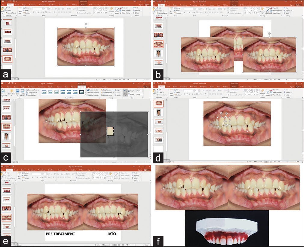
An IVTO was done for a 19-year-old female patient who presented with missing maxillary bilateral lateral incisors and over retained maxillary deciduous canines. Intraoral frontal occlusion photograph of the patient was imported at chairside into the PowerPoint 2016 software. Two copies of the photograph were made followed by cropping of one side maxillary right central incisor and maxillary right canine separately. To replicate the orthodontic treatment plan of orthodontic distalization of the maxillary canines after extraction of deciduous canines and opening space for lateral incisor replacement, the cropped canines were superimposed in a distal position over retained deciduous canines and rotated to match the inclination. The cropped canines from that position were copied and pasted to the other sides with a simple horizontal flip.
For the bilateral lateral incisors replacement, the cropped upper central incisor was shrunk diagonally to replicate the lateral incisors and rotated to mimic lateral incisor angulation. Finally, the cropped image was positioned between upper canines and central incisors. The model wax setup was performed and compared to the generated IVTO [Figure 3].

- Dental IVTO for bilateral missing maxillary lateral incisors with transpositioned canines. (a) Imported Pretreatment image, (b) Image copied and pasted for cropping individual tooth, (c) Maillary central incisor cropped and image resized to resemble lateral incisor, (d) transpositioned maxillary canine cropped and moved distally, (e) Final positioning of cropped images overlapped bilaterally to mimic lateral incisor replacement, (f) Dental IVTO compared with pretreatment and model setup
Discussion
In the digital era, many orthodontists routinely use cephalometric software for case diagnosis, treatment planning, and formulation of computer-generated visual treatment objectives. Morphing and warping of the images have been utilized for predicting orthodontic treatment outcome.[14,15] Now, the commercially available programs allow superimposition of the profile images, use 3D records for simulating treatment outcome in all three dimensions of space, and provide multiple treatment options.[16]
The recent introduction of 3D scanners and cone-beam computed tomography has added to the accuracy of 3D diagnosis and treatment planning in real time. Unfortunately, this technology needs an exorbitant investment and specialized training. An orthodontist has to regularly communicate with a technologist through a remote server to plan the virtual treatment outcome.[17] Generating IVTO in PowerPoint does not require complex technical skills in using imaging software, as a simple diagnostic digital photograph is imported into a more familiar PowerPoint software to create the instant dental and skeletal treatment outcome visualizations which can be shared with the patient at chairside. PowerPoint software comes preinstalled on most computers and can easily be installed in any computer system.
Compared to Microsoft Paint and other photo editing software, PowerPoint offers simplicity and versatility in editing. Exporting images in portable document format, PowerPoint slide show, other image formats, and portability to other devices are easily carried out with the help of PowerPoint. When compared to other commercially available software dedicated to imaging and visual treatment planning, which uses modern-day technology of digital image warping to transform the image pixels to smoothly blend at the edges of the image into a simulated posttreatment image, IVTO made with PowerPoint software produces superimposition images which do not blend in the edges. This does not prove to be a limitation, as the gross nonblended edges in IVTO during superimposition can be precisely blended to smooth contoured image with Photoshop (Adobe Systems Incorporated, Park Avenue, San Jose, CA, USA). However, this might require additional photoshop training. IVTO provides the necessary communication aid in patient education to help patient contemplate dental and skeletal treatment results to match their desire and thus increasing treatment acceptance.
Conclusion
Instant virtual treatment outcome is an efficient, economical, and simple method for generating a precise treatment outcome
A digital photograph can be utilized for generating IVTO, proving its simplicity
Instant chairside visualization for dental and skeletal treatment planning, hence, serves as an excellent patient education tool.
Declaration of patient consent
The authors certify that they have obtained all appropriate patient consent forms. In the form the patient(s) has/have given his/her/their consent for his/her/their images and other clinical information to be reported in the journal. The patients understand that their names and initials will not be published and due efforts will be made to conceal their identity, but anonymity cannot be guaranteed.
Financial support and sponsorship
Nil.
Conflicts of interest
There are no conflicts of interest.
References
- Video Cephalometric Diagnosis (VCD): A new concept in treatment planning? Am J Orthod Dentofacial Orthop. 1996;110:128-36.
- [Google Scholar]
- Treatment planning for orthodontic-restorative cases with sureSmile technology. J Clin Orthod. 2014;48:639-49.
- [Google Scholar]
- Three-dimensional photographs for determining the index of orthodontic treatment need in scientific studies. Am J Orthod Dentofacial Orthop. 2016;150:64-70.
- [CrossRef] [PubMed] [Google Scholar]
- Accuracy of dolphin visual treatment objective (VTO) prediction software on class III patients treated with maxillary advancement and mandibular setback. Prog Orthod. 2016;17:19.
- [Google Scholar]
- Advanced powerPoint animation techniques: Part II. Am J Orthod Dentofacial Orthop. 2000;118:236-40.
- [CrossRef] [PubMed] [Google Scholar]
- Managing digital images. Am J Orthod Dentofacial Orthop. 2000;118:354-8.
- [CrossRef] [PubMed] [Google Scholar]
- Why does the file get too large when I paste a picture in powerPoint? Am J Orthod Dentofacial Orthop. 2004;125:753.
- [Google Scholar]
- Digital photographs and powerpoint. Am J Orthod Dentofacial Orthop. 2004;126:639.
- [CrossRef] [PubMed] [Google Scholar]
- How can I convert my slides to digital images? Am J Orthod Dentofacial Orthop. 2004;126:640.
- [CrossRef] [PubMed] [Google Scholar]
- Inserting a flash movie into a powerPoint presentation. Am J Orthod Dentofacial Orthop. 2007;131:285-7.
- [Google Scholar]
- Use of on-line digital image services to create customized documents for the orthodontic office. Am J Orthod Dentofacial Orthop. 2008;133:771-6.
- [CrossRef] [PubMed] [Google Scholar]
- Powering up your powerPoint presentations. Am J Orthod Dentofacial Orthop. 2000;118:116-20.
- [Google Scholar]
- Morphing and warping. Part I. Am J Orthod Dentofacial Orthop. 1999;115:466-77.
- [CrossRef] [Google Scholar]
- Morphing and warping: Part II. Am J Orthod Dentofacial Orthop. 1999;115:706-8.
- [CrossRef] [Google Scholar]
- Dolphin imaging software: An analysis of the accuracy of cephalometric digitization and orthognathic prediction. Int J Oral Maxillofac Surg. 2005;34:619-26.
- [CrossRef] [Google Scholar]


