

Translate this page into:
Relationship of skeletal malocclusion with eye and hair color in Turkish adolescent patients


*Corresponding author: Taner Ozturk, Department of Orthodontics, Faculty of Dentistry, Erciyes University, Kayseri, Turkey. tanertr35@gmail.com
-
Received: ,
Accepted: ,
How to cite this article: Ozturk T, Ozsaygili C, Topsakal U. Relationship of skeletal malocclusion with eye and hair color in Turkish adolescent patients. APOS Trends Orthod 2021;11(2):148-55.
Abstract
Objectives:
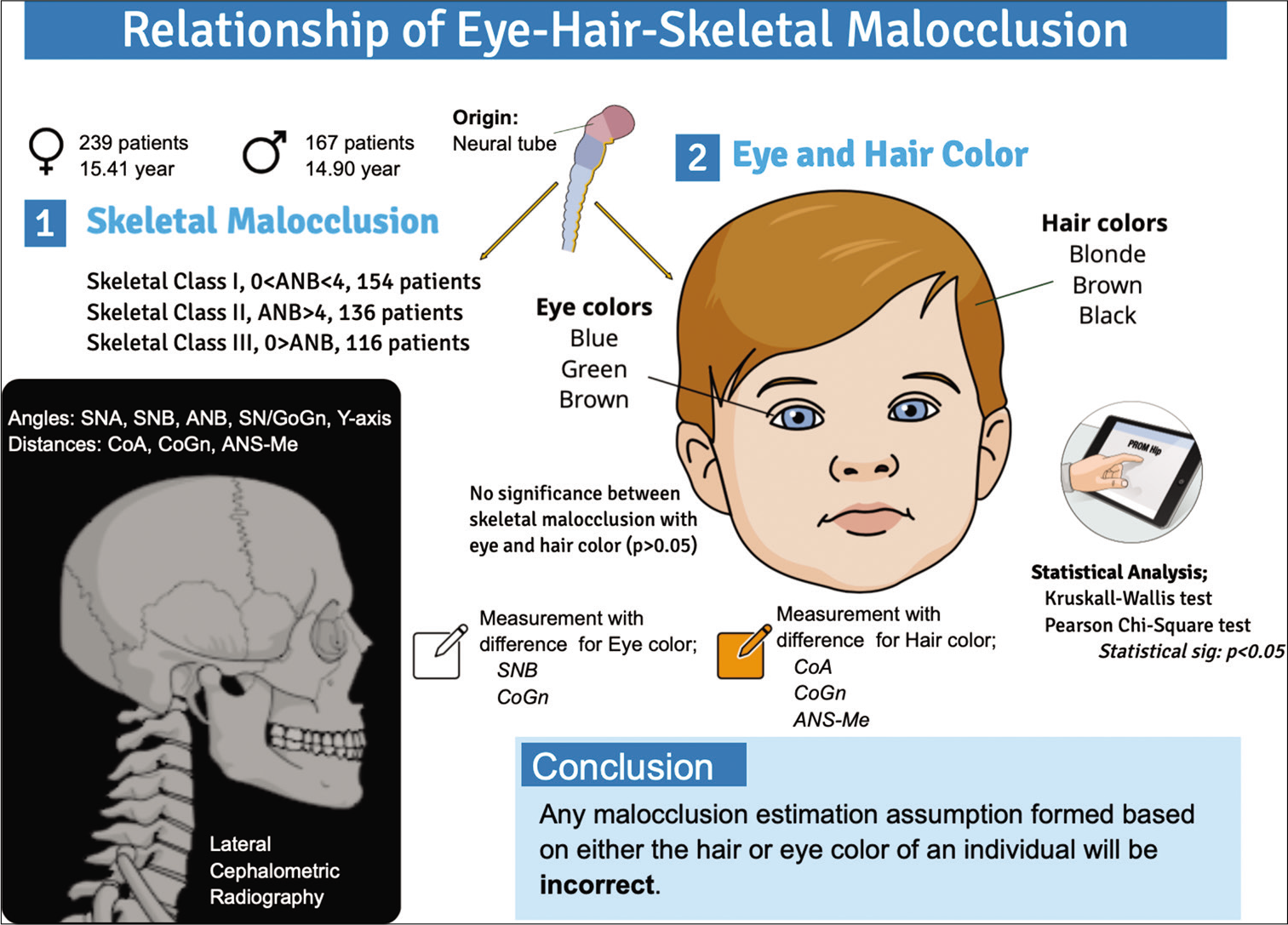
The aim of this study was to establish whether a relationship exists between eye and hair color and orthodontic anomalies; an association has never been evaluated previously.
Materials and Methods:
The records of 406 adolescent patients to the Erciyes University Faculty of Dentistry for orthodontic treatment were included in this retrospective cohort study. Participants were divided into sagittal (Class I, Class II, and Class III) and vertical (low angle, normal angle, and high angle) skeletal malocclusion classes. Moreover, participants were also divided by their eye (brown, green, or blue) and hair (black, brown, or blonde) color. Collated data were statistically evaluated using the SPSS software by applying the one-way analysis of variance, Kruskal–Wallis, the Pearson Chi-square, and Fisher’s exact tests. Statistical significance was accepted at P < 0.05.
Results:
No statistically significant relationships were identified between sagittal and skeletal malocclusion and eye color (P > 0.05). However, the sella-nasion-b and CoGn parameters of brown-eyed individuals were significantly smaller than individuals with other eye colors (P < 0.05). Moreover, a statistically significant difference was established for the CoA, CoGn, and ANS-Me parameters between the different hair groups (P < 0.05). All three parameters were significantly lower in brown-haired individuals compared to individuals with black haired (P < 0.05).
Conclusion:
This study identified no significant association between the eye and hair color variable, with similarly formed craniofacial structures, and with the sagittal and vertical skeletal malocclusion. Therefore, any malocclusion estimation assumption formed based on either the hair or eye color of an individual will be incorrect.
Keywords
Eye color
Hair color
Skeletal malocclusion

INTRODUCTION
During the embryonic period, craniofacial development can be divided into several stages. These stages include the formation of the head plan, the neural tube, and the oropharynx, into the cell migration and interactions required for the formation of craniofacial tissues, the formation of various organ systems (branchial arches, brain, eyes, etc.), and the final stage of tissue differentiation.[1] A study by Kish et al. reported the eye, one of the important sensory structures of the facial region, as one of the structures responsible for the development of craniofacial structures.[2] However, research has established that many syndromic diseases affecting craniofacial structures can affect both the eye and dental structures.[1,2] Based on this information, we can deduce that the different structures in the craniofacial region originate from similar backgrounds. The structures and organs of the craniofacial region that all originate from the same head neural crest cells and from the same dentofacial skeletal features.[1] This deduction is further supported by the knowledge that the melanocytes contributing to both eye and hair color and the chromophore structures that allow the emergence of color in teeth, all have similar origins.[3,4] Furthermore, the functional matrix theory, which was described by Moss, details how the orbital cavity is one of the functional matrices involved in the development of the craniofacial structures and that this structure is closely related to the maxillary bone.[5,6] However, in a study examining the multipotential nature of neural crest cells, it was reported that these cells may be effective in both cartilage and bone tissue formation and melanocyte formation during development.[7] Although the issue of eye and hair color is a matter related to melanocytes,[8,9] it can be assumed that they can be affected by each other and have a relationship since they can be composed of crest cells that take a basic origin.
At present, there are only a few studies in the literature evaluating the relationship between tooth color and/or dental structures and eye or hair color.[10-13] However, there are no studies evaluating the relationship between the phenotypic features of the eye/hair and skeletal malocclusion. This study aimed to assess the relationship between eye and hair color and skeletal malocclusions and to examine the relationship between dentofacial features and eye and hair phenotypic features.
MATERIALS AND METHODS
The Erciyes University Clinical Research Ethics Committee reviewed and approved (Protocol Number: 2020/331) the undertaking of this retrospective cohort study [Figure 1]. According to the power analysis performed before the study, it was determined that a total of 400 subjects should be taken with 0.36 effect size, 5% error, and 90% power. Study participants consisted of 406 adolescent patients (239 females, 15.41 ± 3.26 years; 167 males, 14.90 ± 3.30 years). The patients included in the study were randomly selected among the patients who applied to the Department of Orthodontics, the Faculty of Dentistry, Erciyes University, with the request for orthodontic treatment, according to the presentation date. Inclusion criteria of the study; belonging to the same society (Turkish), not having skin disease affecting the hair and eyes, not having any malformation, trauma, or surgical procedure affecting the craniofacial region, not being artificial of hair color, not using contact lenses, and not receiving orthodontic treatment determined. Extraoral high definition photographs of all participants were taken with a digital camera (Canon, Mark EOS 5D Mark IV, Tokyo, Japan), and eye and hair color assessments were subsequently performed on these photographs. All photographs were taken by the same researcher using a gray background, the same flash system, the same distance, and camera system. Lateral cephalometric radiographs of individuals were taken by the same technician using an Orthoceph cephalometric radiography device (Orthoceph OP300, Instrumentarium, Tuusula, Finland), and skeletal cephalometric analyses were performed with Dolphin Imaging software (version 11.0; Dolphin Imaging and Management Solutions, Chatsworth, USA).
- Brief representation of graphical abstract.
The eye color photographic reference set proposed by Sturm and Larsson was used to assess eye color.[14] This eye color reference set divides eye color groups into three; specifically, blue, green, and brown. Next, the evaluation of hair color was performed using the Bruce Beard’s hair color chart as detailed in Lagouvardos et al.[10] The scale specified in Bruce Beard’s hair color chart divides hair color into three groups: Black, brown, and blonde. For each of the color assessments, three consecutive evaluations were carried out, and two or three identical results were ultimately accepted for the three evaluations. These evaluations were carried out by an ophthalmologist (C.O.), one of the study’s authors.
Craniofacial features were examined using cephalometric analysis [Figure 2a]; including five angular (sella-nasion-a point [SNA]; sella-nasion-b [SNB] point; a point-nasion-b point [ANB]; sella-nasion/gonion-gnathion [SN-GoGn]; and Y-axis [sella-gnathion/Frankfort horizontal line]) and three linear (CoA [maxillary length: Condylion-a point]; CoGn [mandibular length: Condylion-gnathion]; and ANS-Me [anterior nasal spine-menton]) measurements [Figure 2b]. The three skeletal malocclusion classes, as determined by the ANB angle, are described as Classes I (0°<ANB<4°), II (ANB>4°), and III (ANB<0°).[15] The vertical growth pattern classes of the face, according to the SN-GoGn angle, are established as low angle (<26°), normal angle (26°<SN/GoGn<38°), and high angle (>38°).[16] These evaluations were carried out by an orthodontist (T.O.), one of the study’s authors.
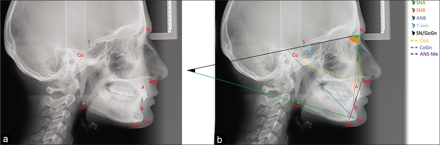
- Representation of cephalometric points and measurements used in the study. (a) Cephalometric points; A: A-point, ANS: Anterior nasal spina, B: B-point, Co: Condylion, Go: Gonion, Gn: Gnathion, Me: Menton, N: Nasion, S: Sella, (b) Cephalometric measurements; SNA: Sella-nasion-A, SNB: Sella-nasion-B, ANB: A-nasion-B, SN/GoGn: Sella-nasion/gonion-gnathion, Y-axis, CoA: Condylion-A, CoGn: Condylion-gnathion, ANS-Me: Anterior nasal spina-menton.
Statistical analysis
Statistical evaluations were conducted using the Statistical Package for the Social Sciences (SPSS, ver. 24.0, IBM, Chicago, USA) software. Before performing the statistical analysis, the normality of the numerical data was assessed using both the Shapiro–Wilk and Kolmogorov–Smirnov tests. Subsequently, normally distributed data were analyzed using the one-way analysis of variance test and all paired comparisons were analyzed using the Fisher’s least significant difference test. In contrast, statistical analysis of non-normally distributed data was performed using the Kruskal–Wallis test and the Mann–Whitney U-test was used to analyze the paired comparisons. All categorical data were analyzed using the Pearson Chi-square test and Fisher’s exact test. Spearman Rho correlation coefficient was used for correlation analysis. Statistical significance was accepted at P < 0.05.
Reliability analysis
To evaluate the reliability of the measurements of the individuals included in the study, the second measurement of 25 randomly selected patients was performed by the same researcher 1 month after the first measurement. Intraclass correlation coefficient (ICC) was used to evaluate the two measurements. Accordingly, the ICC coefficient was determined to be between 0.638 and 0.853.
RESULTS
The results of this cross-sectional study included the assessment of 154 skeletal Class I, 136 skeletal Class II, and 116 skeletal Class III malocclusion individuals [Table 1]. The data indicated no significant relationship between an individual’s eye color and skeletal sagittal malocclusion, vertical growth pattern of the face, and sex [P > 0.05, Table 2]. However, in keeping with the Turkish population, the vast majority of individuals were brown eyed [%70.2, Figures 3a and b]. In addition, it was determined that the majority of brown-eyed patients had a normal angle vertical face pattern [%70.1, Figure 3b]. Evaluation of the cephalometric measurements against eye color established a significant difference in the SNB and CoGn parameters between the three eye groups [P < 0.05, Table 3]. SNB values of blue-eyed individuals were found to be significantly higher than the parameters measured for brown-eyed individuals [P < 0.05, Table 3]. In contrast, the CoGn values of the green-eyed participants were significantly lower than those measured for blue-eyed individuals [P < 0.05, Table 3]. In addition, no statistically significant association was established between an individual’s hair color and their skeletal sagittal malocclusion and their face’s vertical growth pattern [P > 0.05, Table 4]. On the other hand, in keeping with the this population, the vast majority of individuals were black haired [%53.7, Figures 4a and b]. In addition, it has been determined that the majority of individuals with brown (42.9%) and black (53.9%) hair that constitute a significant portion of the population have a normal angle vertical face pattern [Figure 4b]. However, a significant relationship was determined between hair color and gender; with the number of brown-haired females identified as higher than the number of brown-haired males [P < 0.05, Table 4]. Finally, examination of the cephalometric measurements according to the three hair color groups established a significant difference between the CoA, CoGn, and ANS-Me parameters and hair color [P < 0.05, Table 5]. Specifically, brown-haired individuals were found to have significantly lower CoA, CoGn, and ANS-Me values when compared to individuals with black hair. Furthermore, black-haired individuals were found to have significantly higher CoGn values compared to individuals with blonde hair [P < 0.05, Table 5].
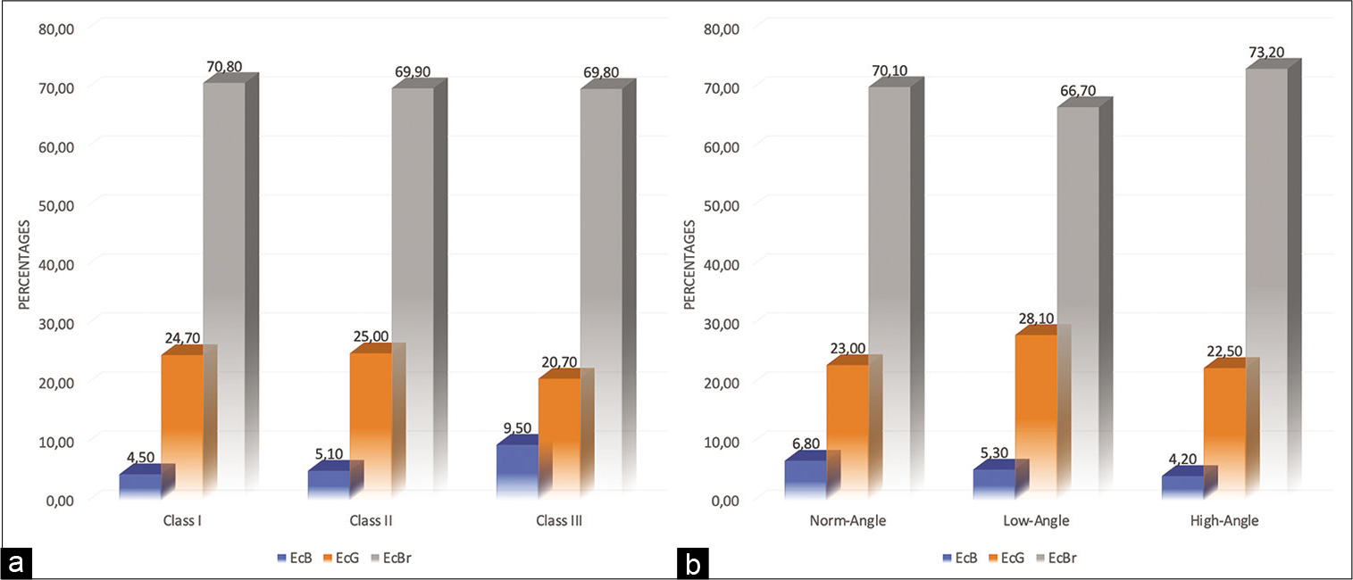
- Distribution of sagittal (a) and vertical (b) skeletal malocclusion classes according to eye color groups. EcB: Eye color blue, EcG: Eye color green, EcBr: Eye color brown.
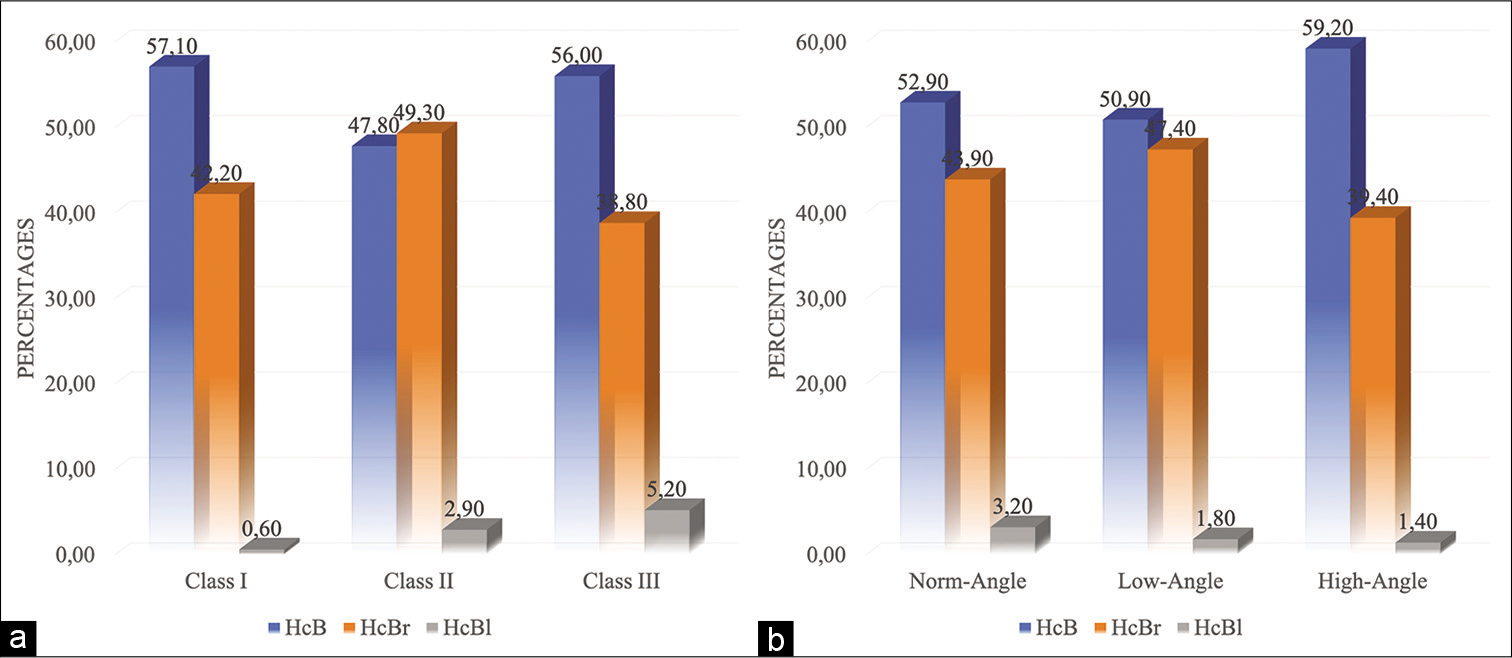
- Distribution of sagittal (a) and vertical (b) skeletal malocclusion classes according to hair color groups. HcB: Hair color Black. HcBr: Hair color Brown. HcBl: Hair color Blonde.
| Females | Males | Totally | |||||||
|---|---|---|---|---|---|---|---|---|---|
| n | % | Age: Mean±SD | n | % | Age: Mean±SD | n | % | Age: Mean±SD | |
| Class I | 96 | 62.34 | 15.18±2.43 | 58 | 37.66 | 15.15±3.14 | 154 | 37.93 | 15.17±2.71 |
| Class II | 89 | 65.44 | 15.37±3.12 | 47 | 34.56 | 14.13±2.45 | 136 | 33.50 | 14.94±2.96 |
| Class III | 54 | 46.55 | 15.91±4.55 | 62 | 53.45 | 15.25±3.90 | 116 | 28.57 | 15.56±4.21 |
| Totally | 239 | 58.87 | 15.41±3.26 | 167 | 41.13 | 14.90±3.30 | 406 | 100.0 | 15.20±3.29 |
n: Number of subject. %: Percentage. SD: Standard deviation
| EcB | EcG | EcBr | Totally | P-values* | |
|---|---|---|---|---|---|
| n(%) | n(%) | n(%) | |||
| Skeletal class | |||||
| Class I | 7 (4.5) | 38 (24.7) | 109 (70.8) | 154 | 0.467 |
| Class II | 7 (5.1) | 34 (25.0) | 95 (69.9) | 136 | |
| Class III | 11 (9.5) | 24 (20.7) | 81 (69.8) | 116 | |
| Vertical growth pattern | |||||
| Normal angle | 19 (6.8) | 64 (23.0) | 195 (70.1) | 278 | 0.834 |
| Low angle | 3 (5.3) | 16 (28.1) | 38 (66.7) | 57 | |
| High angle | 3 (4.2) | 16 (22.5) | 52 (73.2) | 71 | |
| Sex | |||||
| Female | 13 (5.4) | 62 (5.9) | 164 (68.6) | 239 | 0.368 |
| Male | 12 (7.2) | 34 (20.4) | 121 (72.5) | 167 | |
| Totally (n=406) | 25 (6.2) | 96 (23.6) | 285 (70.2) | 406 |
n: Number of subject. %: Percentage of row. Statistical significance P<0.05. EcB: Eye color blue, EcG: Eye color green, EcBr: Eye color brown. *Pearson Chi-square test
| EcB | EcG | EcBr | P-value | Intergroup comparisons, P-value | |||
|---|---|---|---|---|---|---|---|
| Mean±SD | Mean±SD | Mean±SD | EcB-EcG | EcB-EcBr | EcG-EcBr | ||
| SNA | 81.43±4.30 | 80.94±3.90 | 80.33±3.79 | 0.104 | 0.836 | 0.134 | 0.090 |
| SNB | 80.71±5.96 | 78.28±4.47 | 77.95±4.48 | 0.018 | 0.048 | 0.005 | 0.338 |
| ANB | 0.73±4.84 | 2.55±3.18 | 2.38±3.26 | 0.170 | 0.066 | 0.090 | 0.552 |
| SN/GoGn | 32.88±6.25 | 31.91±6.01 | 33.39±6.47 | 0.186 | 0.440 | 0.824 | 0.067 |
| Y-Axis | 59.46±4.50 | 59.42±3.60 | 60.31±5.11 | 0.080 | 0.615 | 0.495 | 0.056 |
| CoA | 79.47±4.69 | 78.61±4.85 | 79.23±5.86 | 0.834 | 0.737 | 0.935 | 0.556 |
| CoGn | 114.97±8.01 | 109.95±7.54 | 111.41±8.56 | 0.032 | 0.009 | 0.056 | 0.139 |
| ANS-Me | 64.35±6.38 | 62.60±5.50 | 63.40±6.51 | 0.326 | 0.156 | 0.407 | 0.275 |
SD: Standard deviation. Statistical significance P<0.05. EcB: Eye color blue, EcG: Eye color green, EcBr: Eye color brown, P values are shown in bold and italics for parameters showing statistical significance
| HcB | HcBr | HcBl | Totally | P-values* | |
|---|---|---|---|---|---|
| n (%) | n (%) | n (%) | |||
| Skeletal class | |||||
| Class I | 88 (57.1) | 65 (42.2) | 1 (0.6) | 154 | 0.089 |
| Class II | 65 (47.8) | 67 (49.3) | 4 (2.9) | 136 | |
| Class III | 65 (56.0) | 45 (38.8) | 6 (5.2) | 116 | |
| Vertical growth pattern | |||||
| Normal angle | 147 (52.9) | 122 (43.9) | 9 (3.2) | 278 | 0.766 |
| Low angle | 29 (50.9) | 27 (47.4) | 1 (1.8) | 57 | |
| High angle | 42 (59.2) | 28 (39.4) | 1 (1.4) | 71 | |
| Sex | |||||
| Female | 112 (46.9)a | 122 (51.0)b | 5 (2.1)a,b | 239 | 0.001 |
| Male | 106 (63.5)a | 55 (32.9)b | 6 (3.6)a,b | 167 | |
| Totally | 218 (53.7) | 177 (43.6) | 11 (2.7) | 406 |
n: Number of subject. %: Percentage of row. Statistical significance P<0.05. HcB: Hair color black, HcBr: Hair color brown, HcBl: Hair color blonde. *Pearson Chi-square test, P values are shown in bold and italics for parameters showing statistical significance
| HcB | HcBr | HcBl | P-value | Intergroup comparisons, P-value | |||
|---|---|---|---|---|---|---|---|
| Mean±SD | Mean±SD | Mean±SD | HcB-HcBr | HcB-HcBl | HcBr-HcBl | ||
| SNA | 80.39±3.92 | 80.67±3.75 | 81.54±3.20 | 0.528 | 0.746 | 0.601 | 0.752 |
| SNB | 78.11±4.80 | 78.17±4.27 | 80.54±6.00 | 0.420 | 0.910 | 0.188 | 0.206 |
| ANB | 2.24±3.46 | 2.50±3.18 | 0.98±4.64 | 0.283 | 0.283 | 0.274 | 0.209 |
| SN/GoGn | 33.29±6.74 | 32.68±5.91 | 32.81±6.26 | 0.844 | 0.566 | 0.863 | 0.911 |
| Y-axis | 60.08±5.28 | 60.09±4.16 | 58.62±3.05 | 0.495 | 0.768 | 0.258 | 0.255 |
| CoA | 79.94±5.72 | 78.14±5.12 | 77.96±7.01 | 0.006 | 0.002 | 0.300 | 0.995 |
| CoGn | 112.68±8.07 | 109.72±8.30 | 108.68±10.34 | 0.001 | 0.001 | 0.048 | 0.519 |
| ANS-Me | 64.07±6.32 | 62.48±6.18 | 60.03±6.28 | 0.009 | 0.031 | 0.090 | 0.414 |
Statistical significance P<0.05. HcB: Hair color black, HcBr: Hair color brown, HcBl: Hair color blonde, P values are shown in bold and italics for parameters showing statistical significance
Significant correlations were found between SNA, SNB, and ANB measurements in all eye color groups [P < 0.05, Table 6]. There are significant correlations between SNA and SN/GoGn, Y-axis, CoA, and CoGn measurements in green- and brown-eyed individuals, unlike blue-eyed individuals [P < 0.05, Table 6]. Significant correlations were found between SNA, SNB, and Y-axis measurements in all eye color groups [P < 0.05, Table 7]. There are significant correlations between SNA and SNB and SN/GoGn, Y-axis, CoA, and CoGn measurements in black- and brown-haired individuals, unlike those with blond hair [P < 0.05, Table 7].
| SNA | SNB | ANB | SN/GoGn | Y Axis | CoA | CoGn | |
|---|---|---|---|---|---|---|---|
| EcB | |||||||
| SNB | 0.478* | ||||||
| ANB | 0.109 | –0.740** | |||||
| SN/GoGn | –0.339 | –0.570** | 0.424* | ||||
| Y Axis | –0.290 | –0.585** | 0.495* | 0.356 | |||
| CoA | 0.135 | –0.069 | 0.190 | –0.356 | –0.006 | ||
| CoGn | –0.028 | 0.590** | –0.735** | –.426* | –0.264 | 0.288 | |
| ANS–Me | –0.404* | –0.296 | 0.018 | 0.120 | 0.529** | 0.097 | 0.400* |
| EcG | |||||||
| SNB | 0.636** | ||||||
| ANB | 0.260* | –0.454** | |||||
| SN/GoGn | –0.468** | –0.415** | –0.011 | ||||
| Y-axis | –0.207* | –0.362** | 0.166 | 0.471** | |||
| CoA | 0.409** | 0.247* | 0.169 | –0.299** | –0.194 | ||
| CoGn | 0.263** | 0.479** | –0.330** | –0.012 | –0.072 | 0.639** | |
| ANS-Me | 0.001 | –0.063 | 0.047 | 0.427** | 0.496** | 0.279** | 0.596** |
| EcBr | |||||||
| SNB | 0.680** | ||||||
| ANB | 0.246** | –0.473** | |||||
| SN/GoGn | –0.455** | –0.556** | 0.214** | ||||
| Y-axis | –0.122* | –0.364** | 0.341** | 0.468** | |||
| CoA | 0.295** | 0.095 | 0.195** | –0.208** | –0.066 | ||
| CoGn | 0.152* | 0.320** | –0.292** | –0.054 | –0.010 | 0.676** | |
| ANS-Me | –0.078 | –0.169** | 0.081 | 0.392** | 0.546** | 0.290** | 0.590** |
The values given are Spearman Rho correlation coefficients. *Correlation is significant at the 0.05 level (two tailed). **Correlation is significant at the 0.01 level (two tailed)
| SNA | SNB | ANB | SN/GoGn | Y Axis | CoA | CoGn | |
|---|---|---|---|---|---|---|---|
| HcBl | |||||||
| SNB | 0.733* | ||||||
| ANB | –0.082 | –0.697* | |||||
| SN/GoGn | –0.445 | –0.528 | 0.191 | ||||
| Y-axis | –0.820** | –0.822** | 0.401 | 0.410 | |||
| CoA | 0.382 | 0.232 | –0.073 | –0.373 | –0.292 | ||
| CoGn | 0.491 | 0.497 | –0.427 | –0.227 | –0.437 | 0.755** | |
| ANS-Me | –0.282 | –0.159 | –0.218 | 0.355 | 0.305 | 0.282 | 0.573 |
| HcBr | |||||||
| SNB | 0.661** | ||||||
| ANB | 0.299** | –0.439** | |||||
| SN/GoGn | –0.437** | –0.508** | 0.133 | ||||
| Y-axis | –0.055 | –0.323** | 0.341** | 0.465** | |||
| CoA | 0.376** | 0.250** | 0.102 | –0.299** | –0.164* | ||
| CoGn | 0.284** | 0.506** | –0.354** | –0.134 | –0.124 | 0.699** | |
| ANS-Me | 0.068 | –0.043 | 0.099 | 0.343** | 0.523** | 0.315** | 0.556** |
| HcB | |||||||
| SNB | 0.674** | ||||||
| ANB | 0.187** | –0.513** | |||||
| SN/GoGn | –0.484** | –0.538** | 0.201** | ||||
| Y-axis | –0.217** | –0.413** | 0.278** | 0.477** | |||
| CoA | 0.254** | 0.019 | 0.252** | –0.179** | 0.011 | ||
| CoGn | 0.085 | 0.289** | –0.318** | –0.004 | 0.081 | 0.602** | |
| ANS-Me | –0.186** | –0.227** | 0.051 | 0.449** | 0.566** | 0.253** | 0.593** |
The values given are Spearman Rho correlation coefficients. HcBl: Hair color blonde, HcBr: Hair color brown, HcB: Hair color black. *Correlation is significant at the 0.05 level (two tailed). **Correlation is significant at the 0.01 level (two tailed)
DISCUSSION
Based on the findings of this study, which determined that there was no significant relationship between eye and hair color and skeletal malocclusion, the null hypothesis was accepted. Nevertheless, the findings also established a difference in certain craniofacial parameters based on the eye and hair color groups. The literature contains several studies that evaluate the relationship between eye structure against various aspects of maxillomandibular structures.[10-13,17-19] However, none of these studies examined the association between eye color and craniofacial skeletal measurements. Although statistical analysis of the collated data did not identify a significant relationship between the color of the eyes and the hair and skeletal malocclusion, the data did determine that certain parameters involved in defining craniofacial skeletal characteristics were significant. Accordingly, although a significant relationship between the sagittal skeletal malocclusion and eye color could not be established, the majority of individuals in this class were brown-eyed, compared to green-eyed individuals who were found to mostly fall under Class III. However, a clinical relationship should not be established between eye color and skeletal malocclusion since the majority of the population has brown eyes.[11] In orthodontic diagnosis and treatment planning, it seems unnecessary to make an additional evaluation according to eye or hair color. However, further studies can be planned using methodologies that will yield different and new findings on large populations.
Furthermore, no significant relationship could be established between eye color and the face’s vertical growth pattern. However, the analysis did establish that the majority of the patients who demonstrated normal growth patterns also had brown eyes. When the position of the mandible relative to the cranial base (SNB) was evaluated, the analyzed data indicated that the mandibular of the blue-eyed individuals was significantly retruded compared to its position in brown-eyed individuals. A study by Alhammadi et al. described the tendency of European individuals to show Class II malocclusions.[20] Therefore, this explains the lower Class II tendency and the lower SNB angle in individuals with brown eyes. Brown eye color was found to be more dominant than the other two eye colors[11] in the population where this study was conducted, as in other European populations. The clinical significance of this indicates that using eye or hair color alone will be insufficient in predicting skeletal malocclusions.
In addition, the research performed by Monaco et al. on the eye did not establish a significant relationship between hyperopia and strabismus, functional problems of the eye, and skeletal malocclusion.[18] These findings also support the findings of this study. In contrast, a different study performed by Monaco et al. reported an association between skeletal malocclusion and vision defects.[21] Furthermore, the study carried out by Lagouvardas et al. did not report a significant relationship between tooth color and eye color.[7] However, evidence has indicated that other characteristics of the facial area are effective in the choice of tooth color. The study by Caruso et al. also reported a significant relationship between molar occlusal and visual defects.[22] In our study, no relationship was observed between eye color and gender. This finding was further supported by the study conducted by Monaco et al. which did not identify a significant relationship between vision defects in the eye and gender.[21] Collectively, all these observations suggest that the structures in the craniofacial region, which originate from similar embryological origins and are located in the same region, interact with each other and may be related to each other.[1]
This study does have its limitations which include the small number of individuals with light-colored eyes and hair color and the small number of samples. However, this study will serve as a guide for further studies.
CONCLUSION
Eye and hair color should not be used as predictors for skeletal malocclusion
There is no significant relationship between sagittal and vertical dentofacial features of the face and eye and hair color, and there is no clinical benefit
Clinically predictable results can be obtained with further studies to be carried out in different populations and large populations.
Statement of ethics
All procedures performed in this study, including data on human participants, were approved by the Erciyes University Clinical Research Ethics Committee (Approval no: 2020/331) and were carried out in accordance with the ethical standards reported in the 1964 Helsinki Declaration.
Declaration of patient consent
Patient’s consent not required as patients identity is not disclosed or compromised.
Financial support and sponsorship
Nil.
Conflicts of interest
There are no conflicts of interest.
References
- Prenatal craniofacial development: New insights on normal and abnormal mechanisms. Crit Rev Oral Biol Med. 1995;6:25-79.
- [CrossRef] [Google Scholar]
- The eye as an organizer of craniofacial development. Genesis. 2011;49:222-30.
- [CrossRef] [PubMed] [Google Scholar]
- Predicting tooth color from facial features and gender: Results from a white elderly cohort. J Prosthet Dent. 2008;99:101-6.
- [CrossRef] [Google Scholar]
- Genetic determinants of hair and eye colours in the Scottish and Danish populations. BMC Genet. 2009;10:88.
- [CrossRef] [PubMed] [Google Scholar]
- Functional cranial analysis of the human maxillary bone: I, basal bone. Angle Orthod. 1967;37:151-64.
- [Google Scholar]
- Craniofacial growth: Evolving paradigms. Cranio. 2015;33:23-31.
- [CrossRef] [PubMed] [Google Scholar]
- Multipotentiality of the neural crest. Curr Opin Genet Dev. 2003;13:529-36.
- [CrossRef] [PubMed] [Google Scholar]
- Hair melanins and hair color: Ultrastructural and biochemical aspects. J Invest Dermatol. 1993;10:S82-9.
- [CrossRef] [PubMed] [Google Scholar]
- Characterization of melanins in human irides and cultured uveal melanocytes from eyes of different colors. Exp Eye Res. 1998;67:293-9.
- [CrossRef] [PubMed] [Google Scholar]
- Tooth, skin, hair and eye colour interrelationships in Greek young adults. Odontology. 2013;101:75-83.
- [CrossRef] [PubMed] [Google Scholar]
- The relationship between tooth color, skin and eye color. Eur Oral Res. 2018;52:50-5.
- [CrossRef] [PubMed] [Google Scholar]
- Relationship of tooth color with skin and eye color based on gender in young Indian adults. Int J Prosthodont Restor Dent. 2019;9:77-81.
- [CrossRef] [Google Scholar]
- Dental occlusion and ophthalmology: A literature review. Open Dent J. 2016;10:460-8.
- [CrossRef] [PubMed] [Google Scholar]
- Genetics of human iris colour and patterns. Pigment Cell Melanoma Res. 2009;22:544-62.
- [CrossRef] [PubMed] [Google Scholar]
- Dehiscence and fenestration in skeletal Class I, II, and III malocclusions assessed with cone-beam computed tomography. Angle Orthod. 2012;82:67-74.
- [CrossRef] [PubMed] [Google Scholar]
- Comparison of pharyngeal airway volume among different vertical skeletal patterns: A cone-beam computed tomography study. Angle Orthod. 2014;84:782-7.
- [CrossRef] [PubMed] [Google Scholar]
- Relationship between mandibular deviation and ocular convergence. J Clin Pediatr Dent. 2004;28:135-8.
- [CrossRef] [PubMed] [Google Scholar]
- Prevalence of hyperopia and strabismus in a paediatric population with malocclusions. Eur J Paediatr Dent. 2011;12:272-4.
- [Google Scholar]
- Correlations between the visual apparatus and dental occlusion: A literature review. Biomed Res Int. 2018;2018:2694517.
- [CrossRef] [PubMed] [Google Scholar]
- Global distribution of malocclusion traits: A systematic review In: Dental Press J Orthod. Vol 23. 2018. p. :40.e1-10.
- [CrossRef] [PubMed] [Google Scholar]
- Prevalence of vision problems in a hospital-based pediatric population with malocclusion. Pediatr Dent. 2013;35:272-4.
- [Google Scholar]
- Association of visual defects and occlusal molar class in children. Biomed Res Int. 2018;2018:7296289.
- [CrossRef] [PubMed] [Google Scholar]


