Translate this page into:
Modified hyrax splint for rapid maxillary expansion in esthetically concerned patients
Address for Correspondence: Dr. Taruna Puri, Bhojia Dental College and Hospital, Baddi, Himachal Pradesh, India. E-mail: tarunapuri5@gmail.com
This article was originally published by Wolters Kluwer and was migrated to Scientific Scholar after the change of Publisher.
Abstract
The orthodontic treatment of Class III malocclusion with a maxillary deficiency is often treated with maxillary protraction either with or without maxillary expansion. The routine procedure for rapid maxillary expansion includes banding on first premolars/first deciduous molars and the permanent first molars. However in some patients who are esthetically very conscious, banding of the first premolar would not be a good esthetic option. So for such circumstances we have designed a modified hyrax splint, which does not need the first premolars to be banded.
Keywords
Modified hyrax splint for rapid maxillary expansion
rapid maxillary expansion
esthetic rapid maxillary expansion splint
INTRODUCTION
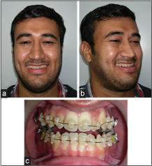
This clinical pearl describes the fabrication of a modified esthetic rapid maxillary expansion (RME) hyrax splint. As the patient who approached to us for orthodontic treatment was esthetic conscious demanded for clear brackets [Damon clear, Figure 1a-c] and he also refused for band formation on first premolar for the fabrication of hyrax splint, and hence we have designed a modified hyrax splint for RME in such esthetically concerned patients.[1]

- (a) The extraoral smiling view, (b) extraoral oblique view, (c) the frontal intraoral photograph
FABRICATION OF THE SPLINT
Banding the first premolar for fabrication of hyrax splint can prove to be esthetically compromising for some patients. So in such cases instead of soldering the wire on a band of first premolar the anterior arm can be bent back and soldered to the first molar. The excess wire from behind the first molar is cut away.
The wire on the lingual surface of first premolar is sandblasted with aluminum oxide particles, and the occlusal surface of first premolar is etched and a piece of super splint [Figure 2] is passed underneath the sandblasted wire [Figures 3 and 4] and it is bonded on occlusal surface of first premolars with flowable composite. In this way, the hyrax wire is connected to the first premolars [Figure 5].
 Figure 2
- The supersplint material
 Figure 3
- The intraoral picture of hyrax splint
 Figure 4
- The supersplint material passed underneath the wire
 Figure 5
- The supersplint bonded to first premolar
Advantages
Easy to fabricate.
As the supersplint connects the hyrax wire to the first premolar, it distributes the forces generated by the appliance evenly to all the teeth.
No need of band formation on upper first premolars.
It can be used in esthetically conscious patients who refuse for a band on first premolar.
The composite placed on the occlusal surface of first premolar will also serve the purpose of a bite block and, therefore, would be beneficial in advancing the maxilla.
Supersplint used in this technique is flexible and has high flexural strength so it won’t get dislodged even with the heavy force application by the hyrax splint. In this case, if the wire would have been bonded directly with composite without using supersplint, the high forces generated by the hyrax splint would have disrupted the bond.
References
- Effects of a modified acrylic bonded rapid maxillary expansion appliance and vertical chin cap on dentofacial structures. Angle Orthod. 2002;72:61-71.
- [Google Scholar]


