Translate this page into:
Roll, pitch, and yaw control using targeted mechanics in clear aligner therapy – A case report

*Corresponding author: Waddah Sabouni, Department of Orthodontic, Bandol Rivage Orthodontie, Sanary-sur-mer, Provence-Alpes-Côte d’Azur, France. wsabounipublications@gmail.com
-
Received: ,
Accepted: ,
How to cite this article: Sabouni W, Al-Ibrahim HM. Roll, pitch, and yaw control using targeted mechanics in clear aligner therapy – A case report. APOS Trends Orthod. doi: 10.25259/APOS_256_2024
Abstract
This case report illustrates the successful use of clear aligner therapy (CAT) in the esthetic correction of a posterior crossbite and crowding in an adult patient. A 33-year-old male presented with a unilateral crossbite on the right side, extending from the first premolar to the second molar, alongside class III molar relationships and bilateral class I canine relationships. The patient exhibited normal overjet, shallow overbite, and mild crowding in both lower and upper dental arches. Traditional fixed appliances were declined in favor of clear aligners. Treatment objectives were focused on correcting the posterior crossbite and achieving an esthetically pleasing outcome with minimal adverse movements. The treatment required correcting malocclusion in all three planes of space - correcting a roll, pitch, and yaw defect by strategic attachment placement, auxiliaries, and careful staging of mesialization and distalization movements. The treatment was completed within 14.5 months, achieving the desired functional and esthetic results. This case demonstrates that clear aligners, when combined with targeted mechanics and staging, can effectively manage complex malocclusions such as posterior crossbite and occlusal cants within a similar timeframe to traditional braces, with a high degree of predictability and patient satisfaction.
Keywords
Clear aligners
Invisalign
Attachment geometry
Staging
Adult orthodontics
3D control
Crossbite
Roll pitch and yaw
Mesialization
Distalization
INTRODUCTION
Clear aligners have become a popular treatment alternative for adult patients seeking an effective and esthetic alternative to traditional orthodontic appliances.[1-3] In addition to the advantages of clear aligners in providing esthetics and comfort to the patients, they enable control of the force system by utilizing planning software and advanced algorithms.[1] However, the orthodontic treatment using clear aligners is not limited to moving teeth digitally.[4] The dental movements resulting from the clear aligners are mechanical movements that release different forces depending on the severity and type of malocclusion, requiring careful pre-planning.[2-4] Improvements and innovations added to the clear aligners, most notably the optimized attachments and customized grading, have increased the precision of tooth movements and provided a wider and more efficient range of orthodontic force delivery.[1,5] Auxiliary methods, such as elastics and partial fixed appliances, contribute to applying adjunctive biomechanics, which lead to more precise control of individual tooth movements and increase the predictability of orthodontic treatment results when using clear aligners.[6]
Ackerman et al. introduced the aeronautical terms “roll, pitch, and yaw” to describe three-dimensional orthodontic problems in spatial planes.[7] With the advent of 3D records, this classification gains importance in analyzing malocclusion deviations across all planes. Much like an airplane, which can move along three planes (front/back, side-to-side, and up/down) and rotate around three axes (horizontal, axial, vertical), the dentition and jaws require a complete description of orientation in space.[7] Roll describes the tipping of the occlusal plane from side to side, pitch refers to its upward or downward tilt in the anterior or posterior regions, and yaw denotes rotation around a vertical axis.[7] These descriptors allow for a more precise analysis of midline deviations, canting, and asymmetries, especially in complex malocclusions involving unilateral Class II or III relationships and crossbites.[7] The inclusion of yaw, previously omitted due to the lack of detection in clinical records, now offers a more comprehensive approach with 3D imaging technologies.[7]
Dentoalveolar expansion is a widely used treatment for maxillary transverse deficiencies with “yaw defects” but must be approached with caution to avoid adverse biological effects.[7-10] Unintended consequences such as gingival recession, alveolar bone deformities (fenestrations and dehiscence), and root resorption have been documented.[8-10] Research suggests that the effectiveness of maxillary expansion decreases as the amount of expansion increases.[10-12] During expansion, posterior teeth undergo both tipping and bodily movement, with tooth inclination increasing proportionally to the degree of expansion.[10-12] Studies also show that many patients develop dehiscences and experience reductions in buccal bone thickness following maxillary expansion.[9,13,14] Therefore, a thorough periodontal evaluation, ideally using cone-beam computed tomography (CBCT), is crucial before initiating treatment.[13,15] CBCT is particularly useful for detecting bone and periodontal defects, especially in adult patients, allowing for better treatment planning and prevention of complications.[13,15]
This article highlights the potential of expansion using clear aligner therapy (CAT) with a precise CBCT evaluation integrated with treatment planning software. In addition, it illustrates satisfying results using an efficacious protocol and precise planning of the staging, the attachments and auxiliary means in correcting a posterior crossbite accompanied by mild crowding in an adult individual.
CASE REPORT
Case history
A 33-year-old male patient came to the clinic with the main complaint of posterior crossbite and crowded anterior teeth. There was no family, genetic, or medical history, and the patient did not undergo any previous orthodontic treatment.
Clinical findings
Extraoral examination revealed facial symmetry with an increased lower facial third in the vertical plane. The upper dental midline coincided with the facial midline, the smile line was high with increased gingival show, and the smile arch was reversed. The profile was concave with a normal clinical the Frankfort-mandibular plane angle (FMA) and retrusive upper and lower lips. On intraoral examination, it was detected a unilateral crossbite on the right side, extending from the first premolar to the second molar, class III molar relationships, class I canine relationships, bilaterally, normal overjet of 1.4 mm; shallow overbite of 1.7 mm; and deviation of the lower dental midline from the upper one by about 2 mm to the right side. Both upper and lower arches were ovoid. Tooth size-arch length discrepancy analysis revealed a mild crowding of 1.5 mm on the upper dental arch and 1.5 mm on the lower dental arch [Figure 1]. Oral health was good, and no parafunctional habits were recorded.
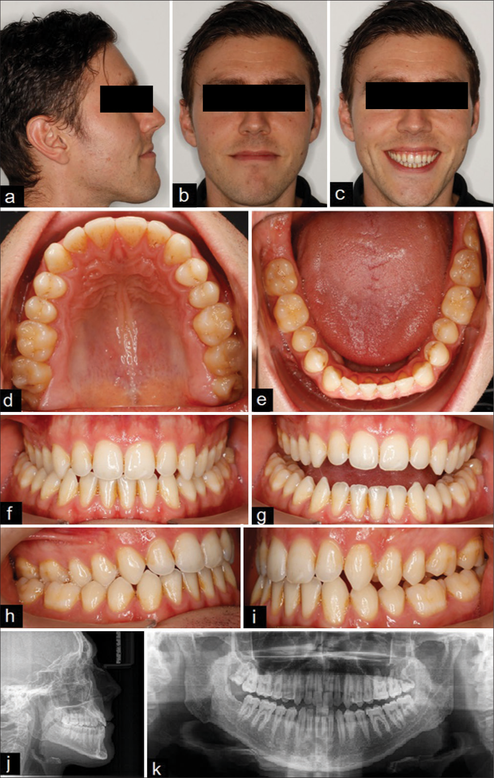
- (a-i) Pre-treatment extraoral and intraoral photos. (j-k) Pre-treatment panoramic radiograph and lateral cephalometric
Radiographic diagnostic assessment
The panoramic radiograph showed full dentition except for the right lower third molar, with no bony or periapical lesions or temporomandibular joint (TMJ) disorders [Figure 1]. Cephalometric analysis [Table 1] demonstrated a skeletal class III pattern with a negative The ANB angle is the difference between SNA (sella-nasion to A point) and SNB (sella-nasion to B point) with a slight vertical growth pattern. Dentally, the upper incisors were normally positioned, while the lower incisors were slightly retroclind with a slightly increased interincisal angle. The CBCT integrated within ClinCheck Pro 6.0 software demonstrated the possibility of posterior dentoalveolar expansion without the formation of fenestration or loss of bone support [Figure 2].
| Parameter | Pre-treatment | Post-treatment | |
|---|---|---|---|
| Skeletal | |||
| 1 | SNA | 73 | 73 |
| 2 | SNB | 75 | 74 |
| 3 | ANB | -2 | -1 |
| 4 | SNPg | 77 | 77 |
| 5 | SN/FH | 11 | 11 |
| 6 | NL/NSL | 8 | 8 |
| 7 | ML/NSL | 38 | 38 |
| 9 | Bjork | 402 | 402 |
| Dental | |||
| 10 | U1-NL- ANGULAR | 103 | 101 |
| 11 | U1-NA- ANGULAR | 26 | 22 |
| 12 | L1-ML- ANGULAR | 92 | 87 |
| 13 | L1-NB-ANGULAR | 29 | 24 |
| 14 | U1/L1 | 127 | 134 |
SNA: the angle between the sella-nasion (SN) plane and the nasion-point.A plane, SNB: the angle between the sella-nasion (SN) plane and nasion-point. B plane, ANB: the difference between SNA and SNB, SNPg: the angle between the sella-nasion (SN) plane and nasion-point. Pog plane, SN/FH: the angle between the sella-nasion (SN) plane and the Frankfort horizontal (FH) plane, NL/NSL: the angle between nasal line (NL) and the cranial base line (NSL), ML/NSL: the angle between the mandibular plane (ML) and the cranial base line (NSL), U1-NL: the angle between the longitudinal axis of the upper incisors and the nasal line (NL), L1-ML: the angle between the longitudinal axis of the lower incisors and mandibular plane (ML), L1-NB: the angle btween the longitudinal axis of the lower incisors and the nasion–point. B line, U1/L1: the angle btween the longitudinal axis of the upper incisors and the longitudinal axis of the lower incisors.
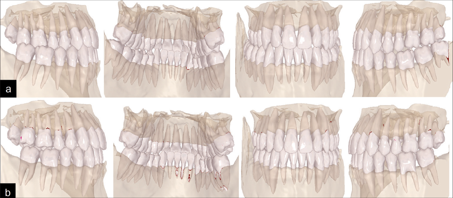
- (a) Pre-expansion assessment using CBCT integrated with ClinCheck. (b) Post-expansion assessment using CBCT integrated with ClinCheck.
Treatment objectives and treatment plan
The treatment objectives included correcting the posterior crossbite, the mild crowding on both arches, the posterior open bite, and the lower midline deviation, and achieving bilateral class I canines and molars relationships with an optimal smile arch and lip line. The preferred treatment method was Invisalign aligners therapy (Align Technology Inc, Santa Clara, CA, USA). It was planned to correct the unilateral crossbite by applying cross-elastics and solve the mild crowding and the lower midline deviation by sequential distalization of the left lower teeth after extraction of the lower left third molar, aided by the application of class III intermaxillary elastics. In addition, the cant correction was planned using intra-maxillary elastics applied between two palatal and buccal mini screws on the left side of the upper jaw. All procedures were carried out in compliance with the ethical principles established in the Declaration of Helsinki.
Therapeutic intervention
Before orthodontic treatment began, the patient was referred to a surgeon to extract the lower left third molar. The treatment consisted of two phases where virtual setup in both occlusion and occlusal views and the tooth movement tables are shown in [Figures 3 and 4]. The virtual setup of ClinCheck Pro 6.0 software in the first phase assumed 42 aligners per arch, where 38 aligners were used for lower molar distalization and upper molar mesialization. In the lower arch, sequential distalization of the left lower teeth and sequential mesialization of the left upper teeth were planned to correct the lower midline deviation and achieve a class I molar relationship and optimal overbite. It was planned to place optimized attachments on most teeth, with vertical 3-mm rectangular ones on the lower left molars and the left upper first molar, to achieve body movements and avoid tipping. The attachments were placed on the labial surfaces of the teeth (upper attachments: #14, #13, #12, #11, #21, #22, #23, #24, #25, #26; lower attachments: #37, #36, #35, #34, #33, #32, #43, #47). 0.2 mm interproximal enamel stripping of the anterior incisors was applied. Metal buttons were placed on the upper teeth (palatally: #16, #15, #14, buccally: #26) and on the buccal surface of the lower teeth (#46, #45, #44) [Figure 3].
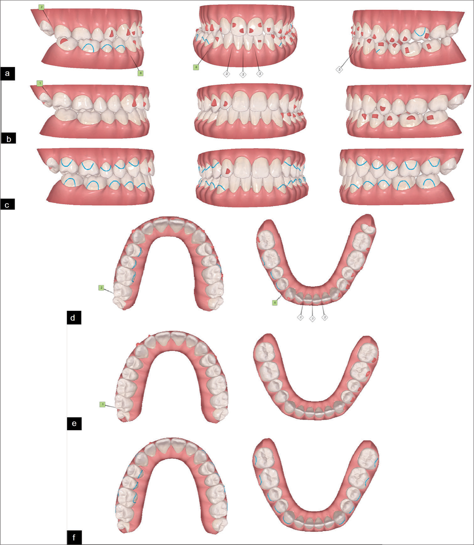
- (a-c) Virtual setup on the ClinCheck™ software (occlusion views): a: at the beginning of the first phase (including 42 aligners; 0.2 mm interproximal enamel stripping between the lower incisors), b: At the beginning of the second phase (including 22 aligners), c: at the end of the treatment. (df) Virtual setup on the ClinCheck™ software (occlusal views): d: at the beginning of the first phase (including 42 aligners; 0.2 mm interproximal enamel stripping between the lower incisors), e: At the beginning of the second phase (including 22 aligners), f: at the end of the treatment.
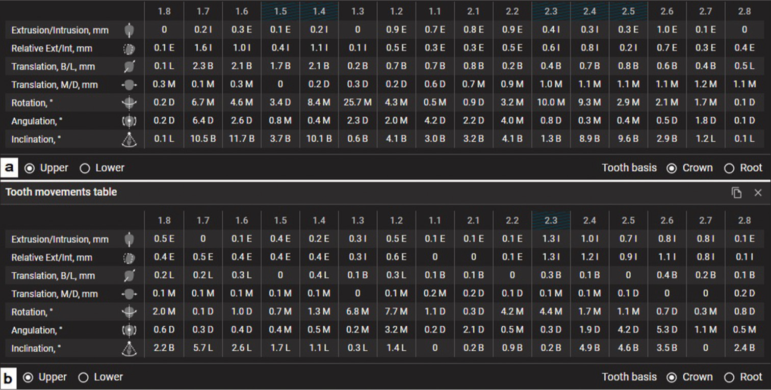
- (a) Tooth movement table on the ClinCheck™ software at the first phase. (b) Tooth movement table on the ClinCheck™ software at the second phase. M/D: Mesial/Distal, B/L: Buccal/Lingual, Ext/Int: Extrusion, Intrusion.
Cross-elastics (1/8 inch, medium force 4.5 oz) were used between each upper tooth (#16, #15, #14) and its corresponding lower tooth (#46, #45, #44) to correct the crossbite. In addition, class III elastics (3/16-inch, medium force 4.5 oz) were applied from a hook on the left lower canine to a button on the buccal surface of the left upper first molar to assist sequential distalization and mesialization movements [Figure 5]. Extraoral and intraoral photographs at the end of the first phase are shown in [Figure 6]. Twenty-two aligners were used in the second phase, where only 8 attachments were placed on the labial surfaces of the teeth (upper attachments: #13, #12, #24; lower attachments: #37, #36, #35, #34, #33) [Figure 3]. Two mini-screws (8 mm × 1.4 mm) were placed between the upper left canine and first premolar buccally and between the upper left first and second premolar palatally, and the patient was asked to apply 1/4 inch, medium force 4.5 oz elastic between the mini-screws to correct the occlusal canting on the left side [Figure 7]. Starting from the 14th aligners, all attachments were removed except for #12, and metal buttons were added to the labial surfaces of the teeth (upper: #17, #16, #14, #24, #25, #26; lower: #37, #36, #35, #34, #44, #46, 47) and only one button was added on the palatal surface of the tooth #27. Vertical elastics (1/8 inch, medium force 4.5 oz) were used between each upper tooth (#17, #16, #14, #24, #25, #26) and its corresponding lower tooth (#36, #35, #34, #44, #46, 47) to settle the bite. In addition, cross-elastics (1/8 inch, medium force 4.5 oz) were used between the palatal button on #27 and the buccal button on #37 to correct the crossbite. The patient was asked to wear the aligners and elastics for 22 h a day and to replace the aligners weekly.
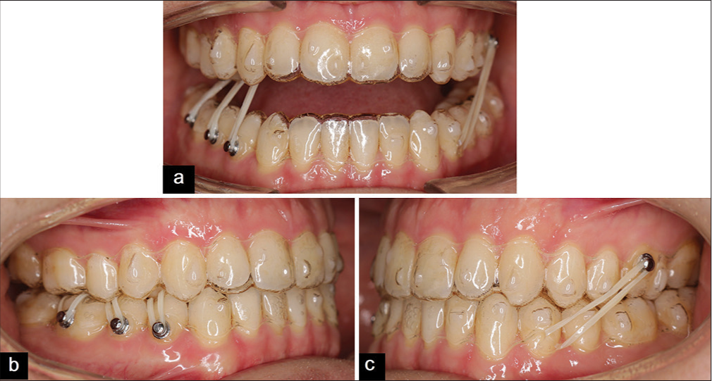
- (a-c) Mid-treatment intraoral photos showing intermaxillary elastics.
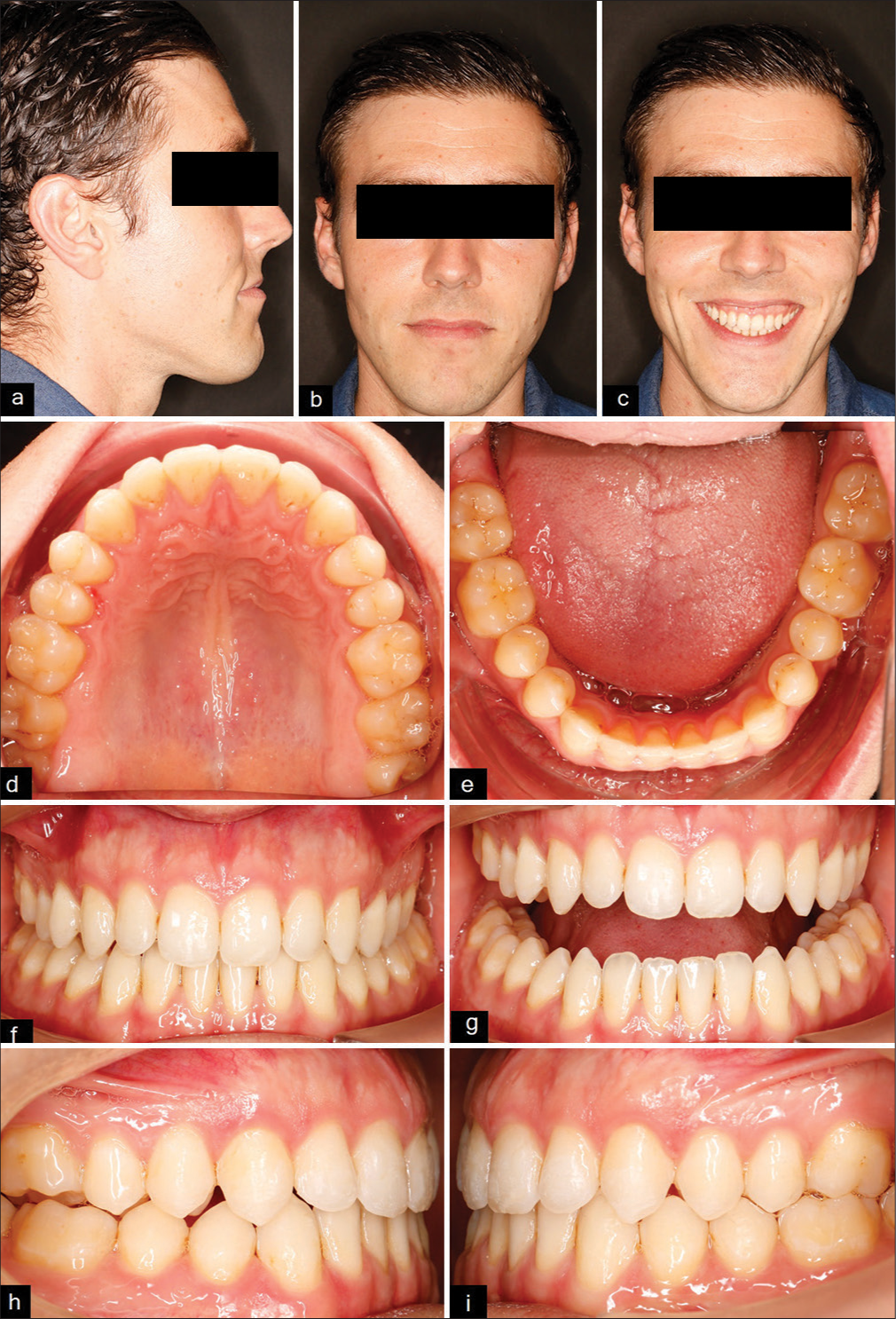
- (a-i) Extraoral and intraoral photos at the end of the first phase.
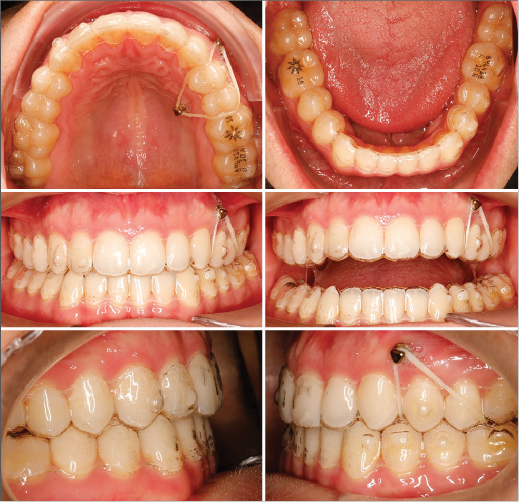
- Mid-treatment intraoral photographs at the second phase showing canting correction.
Treatment outcomes and follow-ups
Overall, treatment time was 14.5 months of active treatment. The treatment was conducted in two phases. Records at the end of the treatment show that the desired objectives have been successfully achieved, as demonstrated in [Figure 8]. The unilateral expansion period was about 9.5 months and was achieved in the first phase, where 42 aligners were used as intended by ClinCheck. The patient’s smile was improved to achieve an ideal smile arc and reduced gingival exposure without occlusal canting. The intraoral records show that the upper and lower dental arches were aligned perfectly with class I relationship bilaterally and no crossbites. The overjet and overbite were satisfactory with coincident dental midlines.
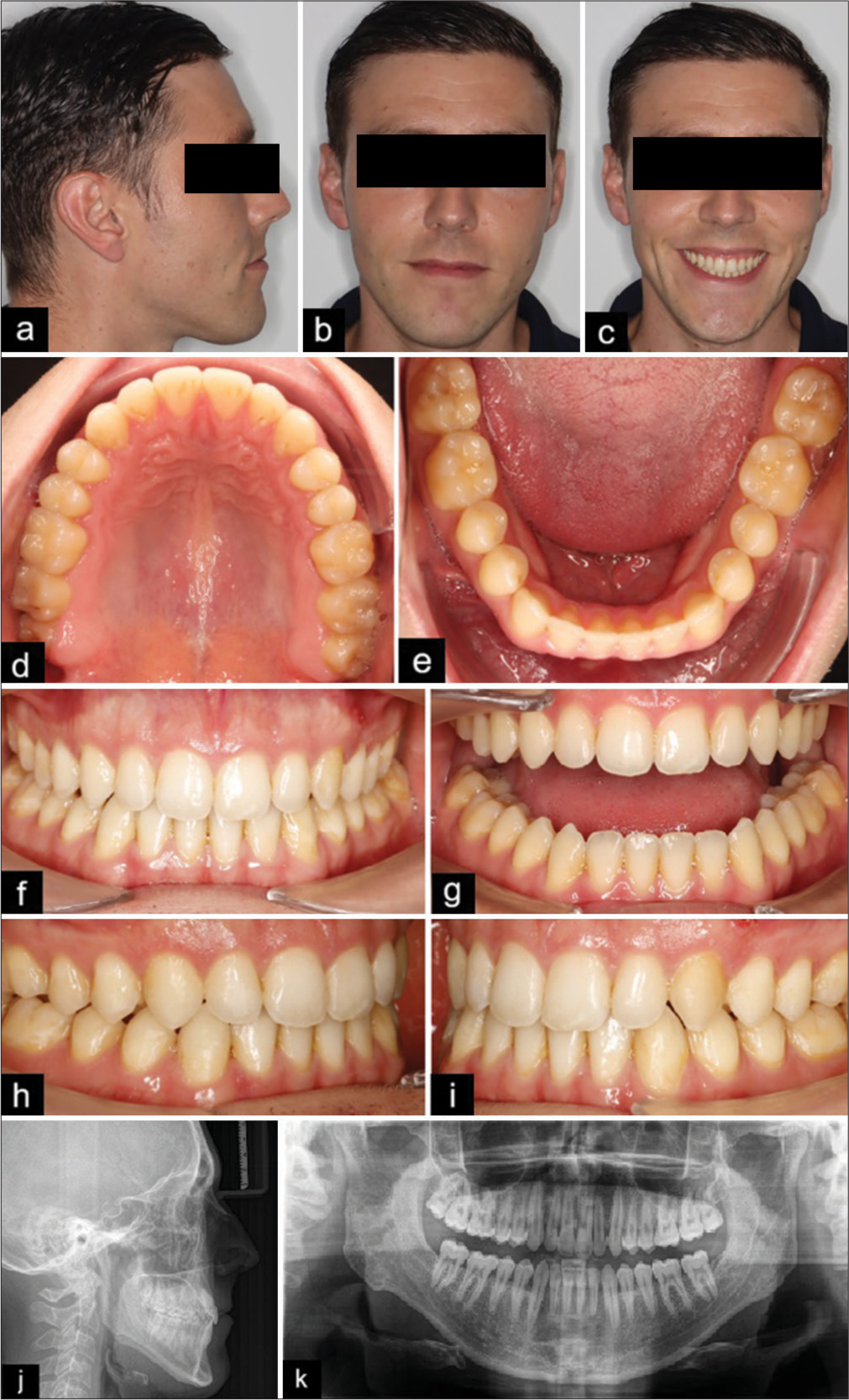
- (a-i) End of treatment records: Extraoral and intraoral photos. (j-k) End of treatment records: Panoramic radiograph and lateral cephalometric radiograph.
The panoramic radiograph at the end of the treatment indicated satisfactory parallelism of the roots without any noted resorption. Digital model and cephalometric superimpositions reveal the uprighting of the lower and upper molars with slight retraction of the lower incisors. There was distalization of the lower left molars with mild extrusion, mesialization of the upper left molars, lingual movements of the lower right posterior teeth, and buccal movement of the upper right posterior teeth due to the application of the cross elastics [Figures 9 and 10]. The patient did not experience or report any negative effects. The patient was very pleased with the result of the treatment and noted improved smile appearance and increased comfort while biting and chewing. The patient was provided with Vivera as removable retainers for retention, and [Figure 11] shows the follow-up intraoral records, which revealed stable treatment outcomes.
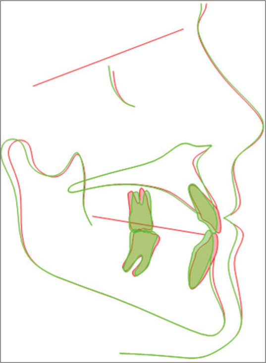
- Superimposition of pre and post lateral cephalometric radiographs. Red pre-treatment and Green post treatment.
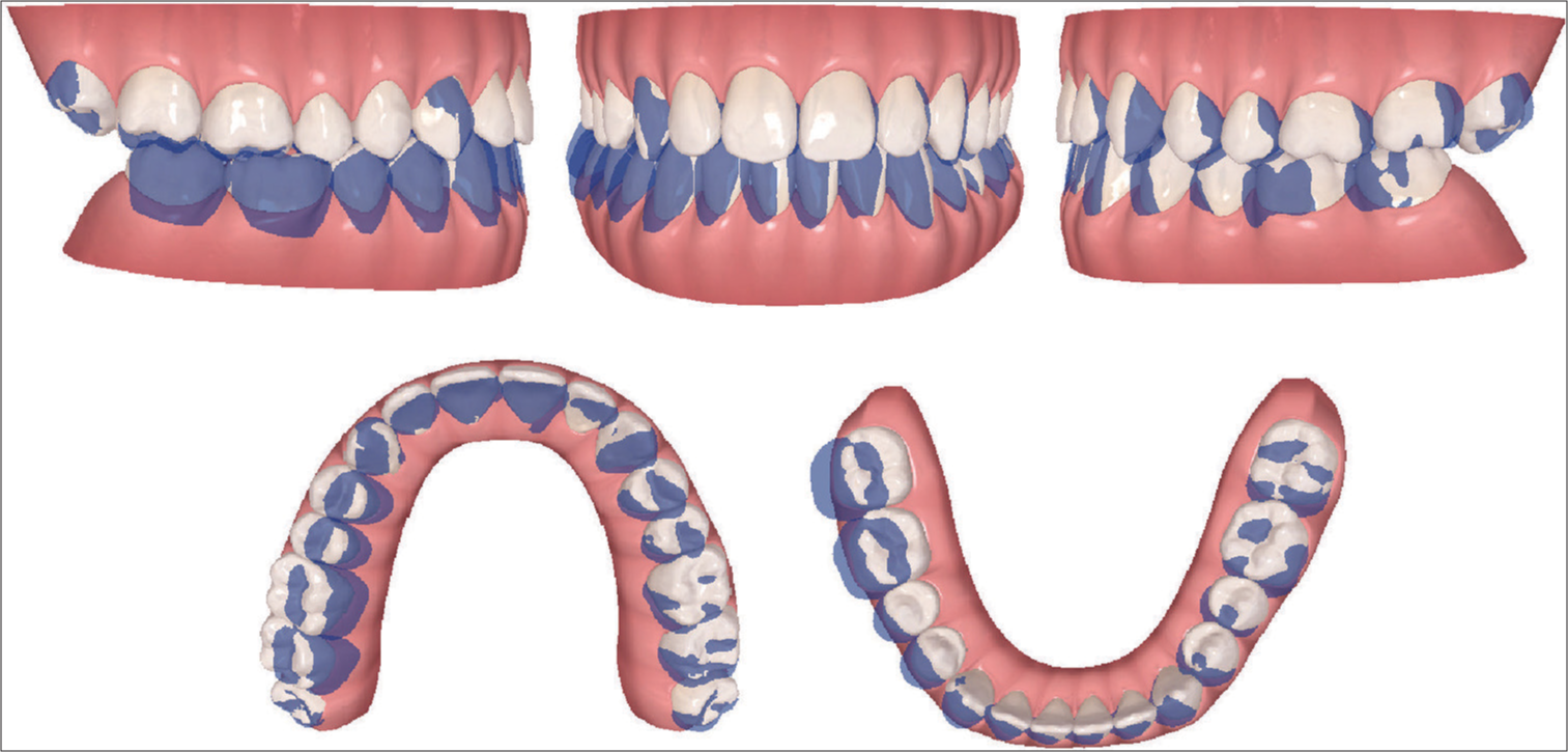
- Superimposition of the digital models at the first phase by the ClinCheck™ software.
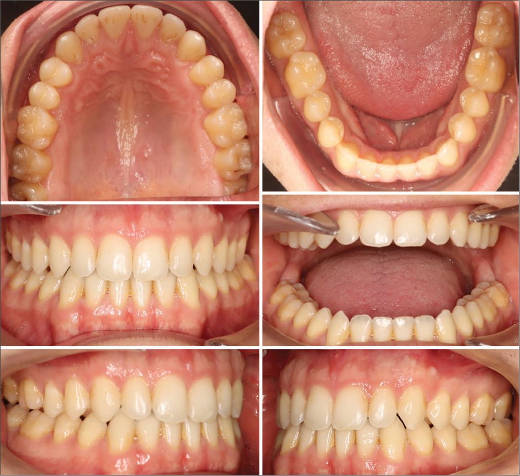
- Intraoral photographs of the follow-up and retention period.
DISCUSSION
The current case report aims to explain the treatment of a “roll, pitch, and a yaw defect” that manifested in a unilateral crossbite, occlusal cant, and a reversed smile line. Intraorally, he presented with dental crowding in an adult patient who opted for clear aligners as an esthetic alternative treatment option to traditional braces. There is still a debate regarding the effectiveness of clear aligners in treating moderate and severe cases of malocclusion. In addition, the number of published papers on the effects of using clear aligners to perform dentoalveolar expansion is still low, as most scientific evidence is focused on the effect of traditional expanders or fixed appliances on the alveolar bone.[16-18]
In adult patients, there are numerous therapeutic techniques have been suggested in scholarly literature for treating crossbites.[10,11,16,17] CAT used for dentoalveolar expansion have been used either alone,[19] with auxiliaries such as intermaxillary elastics,[20] or in combination with some other appliances as a miniscrew-assisted rapid palatal expander.[21] Using intermaxillary elastics in conjunction with clear aligners enabled the correction of unilateral crossbite within a time frame comparable to that of the fixed appliances. This offered the patient an esthetic and comfortable treatment targeted alternative to fixed vestibular appliances.[22]
Clinical trials have shown that using clear aligners in expanding the upper dental arch created buccal tipping of the posterior teeth, extrusion, and lingual tipping of the incisors when expanding the upper dental arch.[10,23,24] The conclusions drawn by Grünheid et al. and Zhou and Guo reported that clear aligners could lead to an increase in interarch width, and the expansion was primarily accomplished through a combination of buccal bodily translation and tipping of the posterior teeth.[10,25] This report aims to underscore the importance of evaluating bone thickness before initiating expansion with clear aligners to prevent alveolar bone fenestrations and dehiscence, which can compromise periodontal health and reduce bony support. The use of CBCT integrated with ClinCheck Pro 6.0 is recommended to assess the feasibility of expansion with Invisalign aligners. In the present case, the buccal inclination of the upper right molars and first premolars ranged from 10° to 11.5°, which was optimal given the initial lingual angulation of these crowns. Conversely, the second upper premolar showed only a 3.7° buccal inclination, while the lower right first and second molars had minimal lingual inclinations of 4.2° and 1°, respectively. This highlights the critical need for pre-treatment adjustment of tooth tip and torque, based on the axis and position of the teeth, before starting orthodontic correction. Notably, no lingual inclination was observed in the upper incisors, likely due to the careful pre-planning of attachment geometry and torque compensation. Specifically, a buccal inclination of 3°–4° was incorporated into the upper incisors’ treatment plan.
According to Morales-Burruezo et al.’s study, dentoalveolar expansion was more effective in the premolar region compared to the second molar region.[20] However, in the current case, expansion proved effective in both the premolar and molar regions, with slightly greater expansion values observed in the second molar region (buccal translation: 2.3 mm, 2.1 mm, 1.7 mm, and 2.1 mm for #17, #16, #15, and #14, respectively) [Figure 2]. This observed variation can potentially be attributed to the fact that 90.4% of the patients included in the Morales-Burruezo et al.[20] study had already exhibited correct positioning of their second molars, rendering the necessity for expansion insignificant.
Distalization conducted in this case was 1.8 mm and 1.6 mm for the second and first lower left molars, respectively. These values are consistent with the average distalization measurements reported in a recent systematic review, which indicated that clear aligners were efficacious in distalizing lower molars by approximately 2–3 mm with high patient compliance rates.[26] Moreover, several articles have emphasized how staging improves the predictability of mesialization and distalization movements.[12,27-29] This strategy was also applied in a recent case to perform sequential mesialization and distalization using 38 aligners to achieve the most predictable movements. In addition, attachments were strategically placed on most teeth to improve movement control and axis alignment, enhancing the predictability of tooth movement. Various attachment designs and angulations were selected, including vertical attachments positioned on the mesial side of the lower left molars to control tipping, prevent mesial rotation, and facilitate bodily movement during distalization.
CONCLUSION
A comprehensive diagnosis is essential when determining the appropriate treatment for maxillary constriction. Evaluating the feasibility of both dentoalveolar and skeletal expansion before treatment is critical. Clear aligners, such as Invisalign, offer a viable option for addressing maxillary dentoalveolar constriction. The integration of CBCT with ClinCheck Pro 6.0 enhances diagnostic accuracy, aiding in precise treatment planning. In this case, the use of aligners demonstrated effective expansion through accurate attachment placement, auxiliary techniques, and careful staging of mesialization and distalization movements. This approach allows for the correction of complex orthodontic issues, including roll, pitch, and yaw deviations, with minimal unwanted tooth movements. Aligners with adjuncts, when smartly planned and staged, provide a predictable and efficient solution for correcting 3D non-skeletal conditions in a time frame that may be comparable to traditional braces. However, further research is necessary to refine our understanding of non-surgical expansion in non-growing adults and improve the predictability of outcomes across all three planes of space.
Data availability statement
The data used to support the findings of this study are included in the article.
Ethical approval
Institutional Review Board approval is not required.
Declaration of patient consent
The authors certify that they have obtained all appropriate patient consent.
Conflicts of interest
There are no conflicts of interest.
Use of artificial intelligence (AI)-assisted technology for manuscript preparation
The authors confirm that there was no use of artificial intelligence (AI)-assisted technology for assisting in the writing or editing of the manuscript, and no images were manipulated using AI.
Financial support and sponsorship
Nil.
References
- An evaluation of the Invisalign® Aligner Technique and consideration of the force system: A systematic review. Syst Rev. 2024;13:43.
- [CrossRef] [PubMed] [Google Scholar]
- Clear aligner therapy concerns: Addressing discrepancies between digitally anticipated outcomes and clinical ground realities. Turk J Orthod. 2024;37:130-9.
- [CrossRef] [PubMed] [Google Scholar]
- Biomechanics of clear aligners: Hidden truths & first principles. J World Fed Orthod. 2022;11:12-21.
- [CrossRef] [PubMed] [Google Scholar]
- Has Invisalign improved? A prospective follow-up study on the efficacy of tooth movement with Invisalign. Am J Orthod Dentofacial Orthop. 2020;158:420-5.
- [CrossRef] [PubMed] [Google Scholar]
- Customized adjuncts with clear aligner therapy: “The Golden Circle Model” explained! J World Feder Orthod. 2022;11:216-25.
- [CrossRef] [PubMed] [Google Scholar]
- Pitch, roll, and yaw: Describing the spatial orientation of dentofacial traits. Am J Orthod Dentofacial Orthop. 2007;131:305-10.
- [CrossRef] [PubMed] [Google Scholar]
- Comparison of alveolar bone thickness and height after slow expansion using quad-helix or clear aligners. Saudi Dent J. 2023;35:255-62.
- [CrossRef] [PubMed] [Google Scholar]
- Maxillary alveolar bone evaluation following dentoalveolar expansion with clear aligners in adults: A cone-beam computed tomography study. Korean J Orthod. 2023;53:264-75.
- [CrossRef] [PubMed] [Google Scholar]
- Efficiency of upper arch expansion with the Invisalign system. Angle Orthod. 2020;90:23-30.
- [CrossRef] [PubMed] [Google Scholar]
- Maxillary arch development with Invisalign system. Angle Orthod. 2021;91:433-40.
- [CrossRef] [PubMed] [Google Scholar]
- Staging of orthodontic tooth movement in clear aligner treatment: Macro-staging and micro-staging-a narrative review. Appl Sci. 2024;14:6690.
- [CrossRef] [Google Scholar]
- Evaluation of maxillary buccal alveolar bone before and after orthodontic alignment without extractions: A cone beam computed tomographic study. Angle Orthod. 2018;88:748-56.
- [CrossRef] [PubMed] [Google Scholar]
- Assessment of posterior dentoalveolar expansion with Invisalign in adult patients. Int J Environ Res Public Health. 2023;20:4318.
- [CrossRef] [PubMed] [Google Scholar]
- A cone-beam computed tomographic evaluation of alveolar bone dimensional changes and the periodontal limits of mandibular incisor advancement in skeletal Class II patients. Angle Orthod. 2020;90:330-8.
- [CrossRef] [PubMed] [Google Scholar]
- Comparison of different types of palatal expanders: Scoping review. Children (Basel). 2023;10:1258.
- [CrossRef] [PubMed] [Google Scholar]
- A comparison of treatment effectiveness between clear aligner and fixed appliance therapies. BMC Oral Health. 2019;19:24.
- [CrossRef] [PubMed] [Google Scholar]
- Maxillary expansion: A comparison of damon self-ligating bracket therapy with MARPE and PAOO. Biomed Res Int. 2022;2022:1974467.
- [CrossRef] [PubMed] [Google Scholar]
- Efficacy and accuracy of maxillary arch expansion with clear aligner treatment. Int J Environ Res Public Health. 2023;20:4634.
- [CrossRef] [PubMed] [Google Scholar]
- Arch expansion with the Invisalign system: Efficacy and predictability. PLoS One. 2020;15:e0242979.
- [CrossRef] [PubMed] [Google Scholar]
- Class III malocclusion and bilateral cross-bite in an adult patient treated with miniscrew-assisted rapid palatal expander and aligners. Angle Orthod. 2018;88:649-64.
- [CrossRef] [PubMed] [Google Scholar]
- Incognito™ lite lingual orthodontic appliance: enhancing the scope of targeted mechanics with customized computer-aided design/computer-aided manufacturing therapy. APOS Trends Orthod. 2015;5:120-3.
- [CrossRef] [Google Scholar]
- The finite element method: A tool to study orthodontic tooth movement. J Dent Res. 2005;84:428-33.
- [CrossRef] [PubMed] [Google Scholar]
- Effects of upper arch expansion using clear aligners on different stride and torque: A three-dimensional finite element analysis. BMC Oral Health. 2023;23:891.
- [CrossRef] [PubMed] [Google Scholar]
- How accurate is Invisalign in nonextraction cases? Are predicted tooth positions achieved? Angle Orthod. 2017;87:809-15.
- [CrossRef] [PubMed] [Google Scholar]
- Efficacy of clear aligners in treating class III malocclusion with mandibular molar distalization: A systematic review. Cureus. 2023;15:e48134.
- [CrossRef] [Google Scholar]
- Staging orthodontic aligners for complex orthodontic tooth movement. Turk J Orthod. 2021;34:202-6.
- [CrossRef] [PubMed] [Google Scholar]
- Lower molar distalization using clear aligners: Bodily movement or uprighting? A preliminary study. Appl Sci. 2022;12:7123.
- [CrossRef] [Google Scholar]
- Accelerated Invisalign treatment of patients with a skeletal Class III. J Aligner Orthod. 2017;1:37-57.
- [Google Scholar]


