

Translate this page into:
A modified dis-impaction spring for impacted canines
Address for Correspondence: Dr. Syed Omar Aziz Rizvi, Vydehi Institute of Dental Sciences and Research Center, #82 EPIP Area, narullahalli, Whitefield, Bangalore, Karantaka, India - 560 066. E-mail: rizvi.omar@gmail.com
This article was originally published by Wolters Kluwer and was migrated to Scientific Scholar after the change of Publisher.
Abstract
Tooth impaction is the retardation in the eruption pattern of a tooth. One of the most commonly impacted teeth is the maxillary canine. However, impaction of mandibular canines is not as common as maxillary canines. Treatment of such impacted teeth usually involves surgical exposure, followed by bonding of an orthodontic attachment to facilitate extrusive movement of the impacted tooth. However, some side-effects on other teeth can be expected which includes the intrusion and tipping of adjacent teeth. In order to prevent side-effects on the adjacent teeth, we present a modified uprighting spring used to extrude an impacted canine.
Keywords
Dis-impaction spring
extrusive movement
impacted canine
uprighting spring

INTRODUCTION
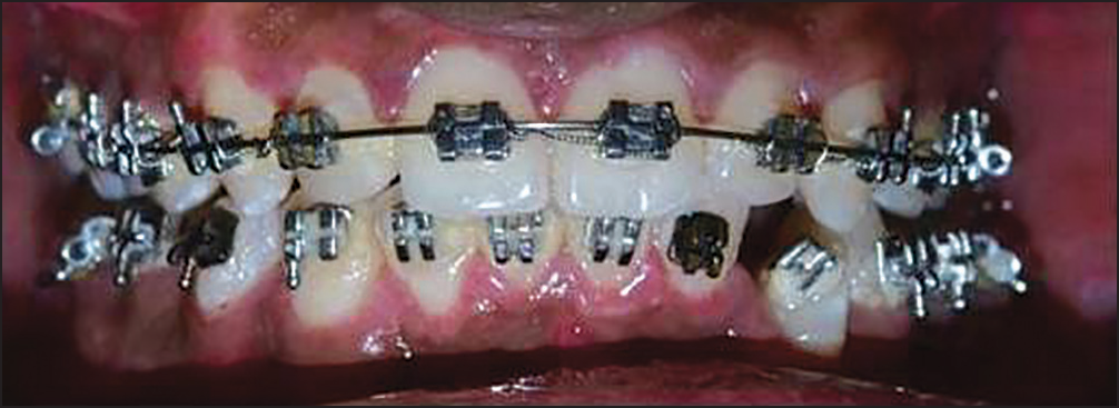
Tooth impaction is the retardation in the eruption pattern of a tooth. One of the most commonly impacted teeth is the maxillary canine.[1] However, impaction of mandibular canines is not as common as maxillary canines [Figure 1].

- Impaction of mandibular canine (33)
Treatment of impacted teeth usually involves surgical exposure followed by bonding of an orthodontic attachment to facilitate extrusive movement of the impacted tooth. Various methods of canine disimpaction have been presented like autotransplantation of the canine, ballista spring system, multiple eyelet chain, tunnel-traction, etc. However some side effects on other teeth, which includes the intrusion and tipping of adjacent teeth, can be expected.[2] In order to prevent side effects on the adjacent teeth, we present a modified uprighting spring used to disimpact and extrude an impacted canine.
DESIGN AND BIOMECHANICS
The design of the spring is similar to that of a Begg uprighting spring.[3] The spring consists of an active arm, a coil, and a stem. The design was modified by making the active arm and helix perpendicular to the stem [Figures 2 and 3]. It is made out of 0.016” AJ Wilcock (premium plus) steel wire. Advantage of using 0.016” AJ Wilcock steel wire with helix is that a constant force within physiological limits is applied.
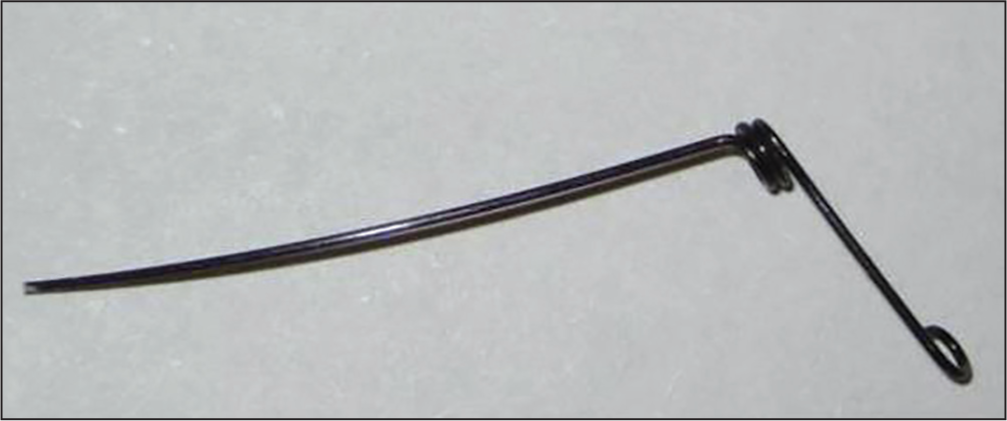
- Modified disimpaction spring
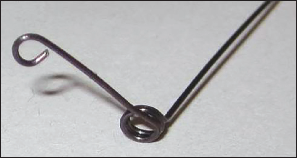
- Modified disimpaction spring
The resiliency of the 0.016” AJ Wilcock wire and the elasticity added by the spring provide an extrusive force on the impacted tooth while the spring unwinds to its passive state [Figure 4]. Reactivation of the spring can be done by making a gingivally directed V bend in the active arm of the spring using an optic plier. This will elevate the hook of the active arm further away from the impacted tooth.
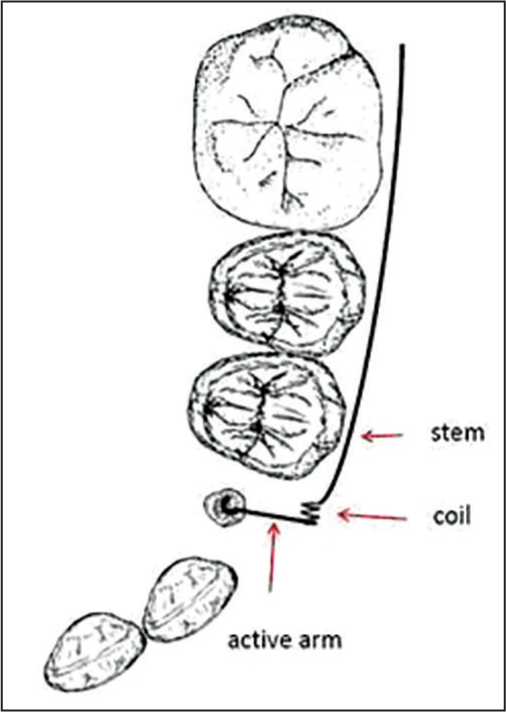
- Schematic representation of the biomechanics of the spring
The force required for the extrusive movement of an impacted tooth is in the range of 60-75 g, which is achieved using this spring.
Only a vertical eruptive movement is desired with this spring without any moment generation. This is achieved by a single point contact while tying the spring to the tooth. If desired, one can shorten the active arm to deliver the same.
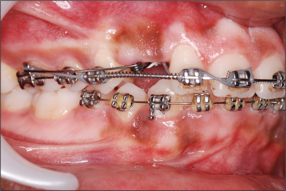
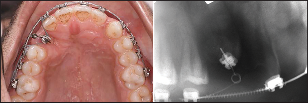
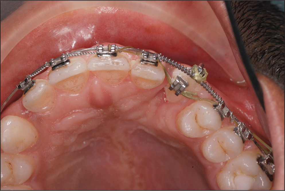
The size of the helix is 2 mm having 2½ turns of wire [Figure 3]. The length of the spring’s stem is such that it extends into the auxiliary tube on the first permanent molar and is tied into the premolar brackets in a piggyback fashion over stainless steel base archwire. This prevents any unwanted movement of the adjacent premolars. A hook is made at the end of the active arm which is pointing toward the upper arch in the passive state. The active arm is then pulled down and engaged in the attachment on the canine thus delivering a constant eruptive force [Figures 5-7]. Length of the active arm is gradually reduced as the canine erupts.
- Pretreatment
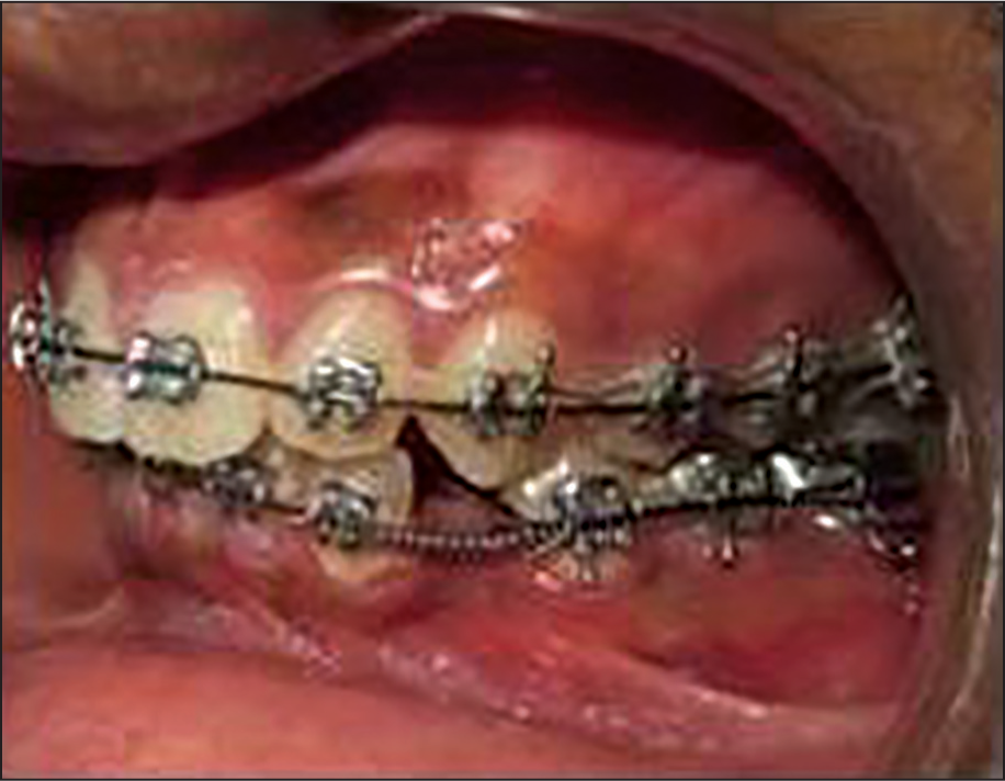
- Spring engaged
- Lateral view of the spring
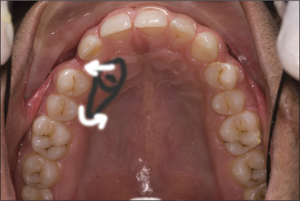
The pictures illustrate the use of this dis-impaction spring for an impacted mandibular left canine. The spring was re-activated every 6 weeks. Re-activation is by opening the helix to adjust the extrusive force on the impacted canine. This modified spring brought about extrusion of the impacted canine over a period of 8 months [Figures 8-10].
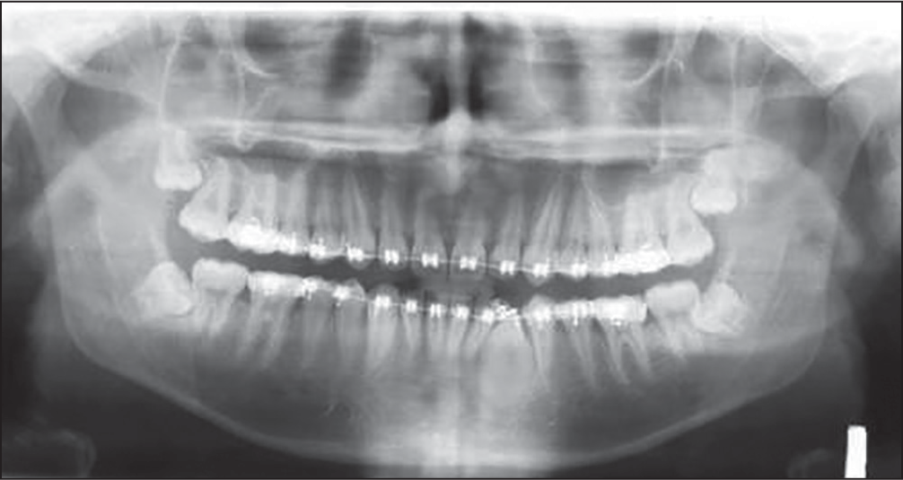
- Orthopantomogram showing the spring in place with canine disimpacted
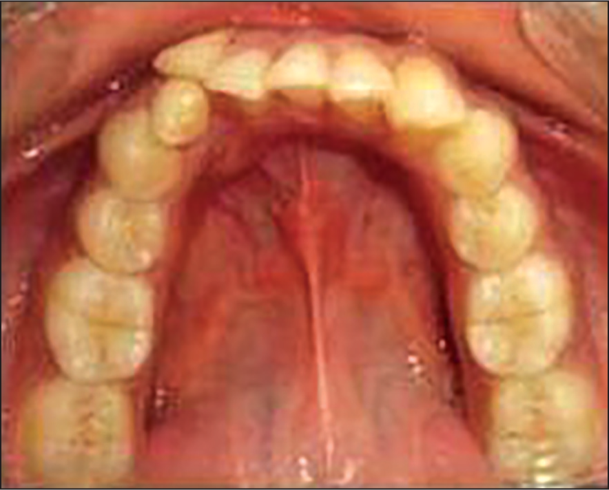
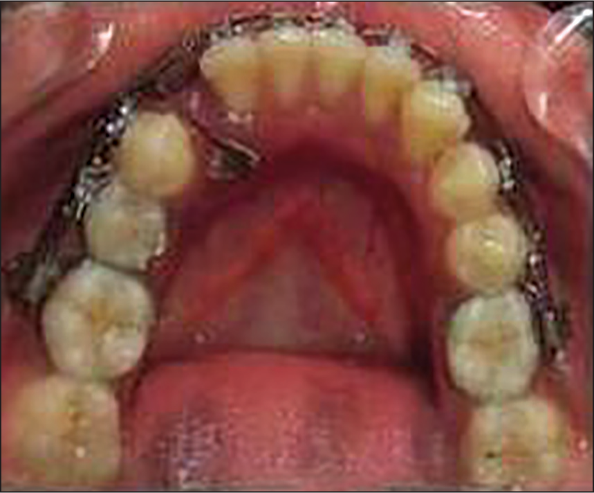
- Dis-impaction of canine completed (occlusal aspect)
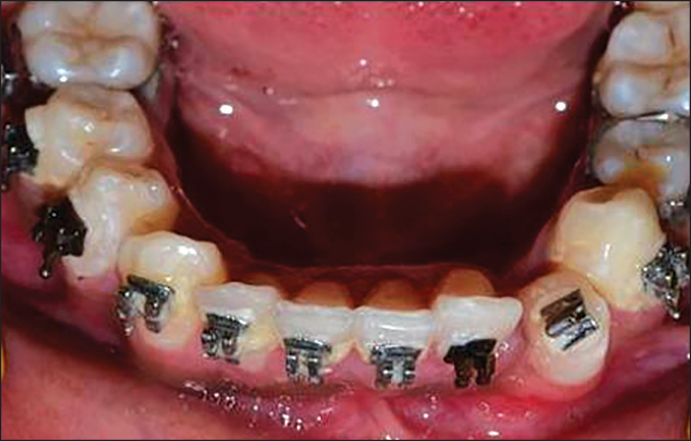
- Dis-impaction of canine completed (frontal aspect)
APPLICATION OF MODIFIED UPRIGHTING SPRING FOR UPRIGHTING A PALATALLY IMPACTED CANINE
The modified spring can also be used to upright the impacted canine while assisting in its eruption. This is depicted in the following case.
A palatal impaction was seen in this case with its crown near the roots of the lateral incisor and the root extending palatal and superior to the roots of the first and second premolars as shown in [Figure 11] with its palatal surface right below the mucosa.
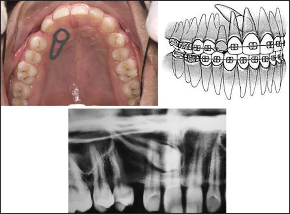
- Location of impacted canine
The spring that was engaged in the vertical space between the wings of the bracket was activated by engaging the spring hook into the hook of the first molar tube [Figures 12 and 13]. The canine was exposed first time by raising a flap, and an indirect pull was applied through a ligature passing through a Begg bracket bonded to the tooth. The second time it was exposed by a palatal window [Figure 14]. The duration of 6 months was taken for the disimpaction, buccal traction and simultaneous uprighting of the canine tooth with the modified spring. Subsequent to the exposure, by attaching a zero degree tip lower incisor bracket to the palatal surface of the canine and by buccal traction with an echain and simultaneous root uprighting, the canine moved to a position shown in Figure 15.
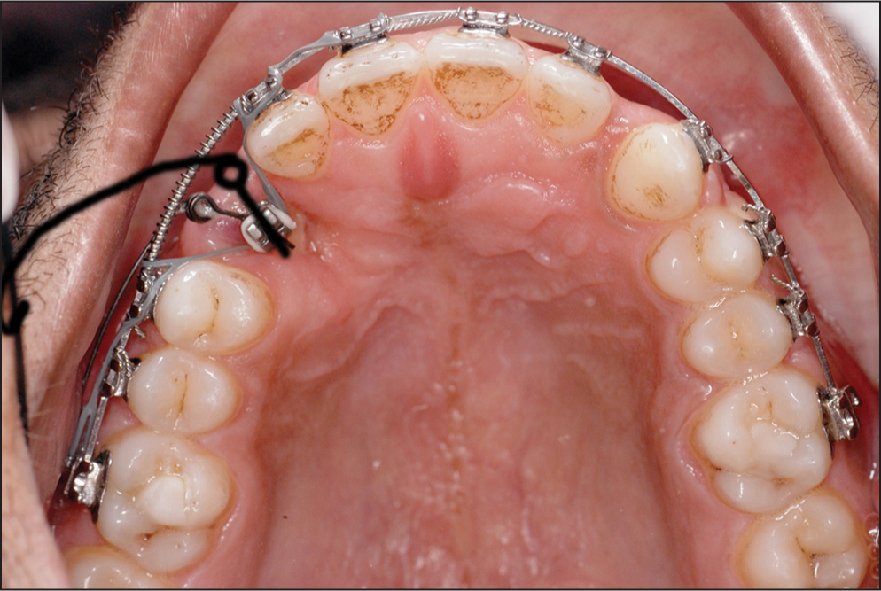
- An uprighting spring (passive position indicated by the black drawing) that delivered approx. 100 g force for uprighting the canine root
- The uprighting spring in activated position with its hook engaged in hook of the molar tube
- Canine position after 6 months of traction and simultaneous uprighting. Note the significant uprighting of the root achieved with the uprighting spring as seen by the alignment of the bracket
- Buccal emergence of the canine. Approximately, a year of traction finally made the canine emerge from the buccal gingiva as seen palatally
BIOMECHANICS
The mechanics [Figure 16] involved a buccal pull on the crown through the first exposure to take it away from the root of the lateral incisor into the space between the crowns of the lateral incisor and first premolar. Once this was achieved, the modified spring was used to upright the root till the root lined up parallel to the premolar roots mesiodistally [Figure 14]. In passive position, the uprighting spring had a position as shown by the black drawing [Figure 12]. The spring was engaged in the vertical space between the wings of the bracket and was secured by either an elastic or metal ligature. It was activated by engaging the spring hook into the hook of the first molar tube.
- Intended mechanics of canine traction
CONCLUSION
This case report has demonstrated the use of modified uprighting springs to dis-impact and extrude and upright impacted canines.
References
- The Orthodontic Treatment of Impacted Teeth (2nd ed). London: Informa Healthcare; 2007. p. :5.
- Extrusion of impacted teeth using mini-implant mechanics. J Clin Orthod. 2012;46:150-5.
- [Google Scholar]
- Refined Begg for Modern Times. India: Mrs Anuradha V Jayde Eureka Towers; 2001. p. :100-2.


