Generic selectors
Exact matches only
Search in title
Search in content
Post Type Selectors
Filter by Categories
Book Review
Case Report
Case Series
Clinical Article
Clinical Innovation
Clinical Pearl
Clinical Pearls
Clinical Showcase
Clinical Technique
Critical Review
Editorial
Expert Corner
Experts Corner
Featured Case Report
Guest Editorial
Letter to Editor
Media and News
Orginal Article
Original Article
Original Research
Research Gallery
Review Article
Special Article
Special Feature
Systematic Review
Systematic Review and Meta-analysis
The Experts Corner
View/Download PDF

Translate this page into:

Clinical Technique
7 (
1
); 52-53
doi:
10.4103/2321-1407.199177

Modified laceback for canine retraction

Departments of Orthodontics, Chandra Dental College, Barabanki, Uttar Pradesh, India
Departments of Pedodontics, Chandra Dental College, Barabanki, Uttar Pradesh, India
Department of Orthodontics, The Oxford Dental College, Bengaluru, Karnataka, India
Department of Orthodontics, Dasmesh Institute of Research and Dental Sciences, Faridkot, Punjab, India

Address for Correspondence: Dr. Stephen Chain, Department of Orthodontics, Chandra Dental College, Barabanki, Uttar Pradesh, India. E-mail: steve911us@gmail.com

Licence
This is an open access article distributed under the terms of the Creative Commons Attribution-Non Commercial-Share Alike 3.0 License, which allows others to remix, tweak, and build upon the work non-commercially, as long as the author is credited and the new creations are licensed under the identical terms.
Disclaimer:
This article was originally published by Wolters Kluwer and was migrated to Scientific Scholar after the change of Publisher.

Abstract

This is a modified lace back to distalize canine. This modification overcomes the previous problems associated with the design.

Keywords

Canine
laceback
open coil

INTRODUCTION

The use of elastic has been used aplenty in orthodontics. They are beinig used in the actice ligation, active tie back and most commonly in the closure of spaces. The use of elastomeric modules as an adjunct to the laceback has been described in Systemized Orthodontic Treatment Mechanics.[1] This is to retract the canine and make space for the blocked lateral or to upright a tipped canine. The use of elastomeric comes with its demerits such as:

  • Increased amount of force

  • Rapid force degradation

  • Appliance removal while reactivating.

To overcome these problems we have devised a technique of active lace back using an open coil spring (3 mm × 020”) with 0.009” stainless steel ligatures (3M Unitek, Monrovia, CA 91016, USA). The ligature is placed on the brackets as the laceback with the ends crossed mesial to the canine before placing the arch wire [Figure 1]. An open coil spring of 8 mm is placed in the wire and the archwire is placed in the brackets and the canine bracket is ligated with the metal ligatures [Figure 2]. Mesial to the canine the open ends of the ligatures are brought forward and the coil spring is compressed with the ligature winding it onto the wire and closing the spring [Figure 3]. When the open coil spring unwinds itself it pushes the canine distally.
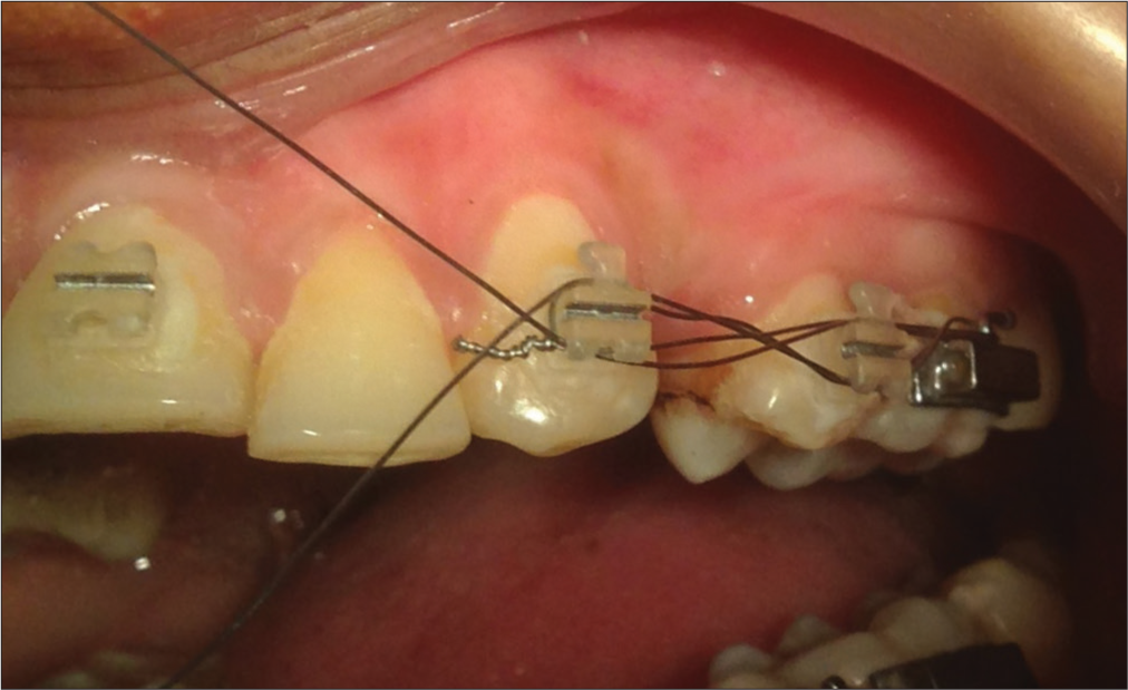
The ligature wire is placed without the placement of the arch wire
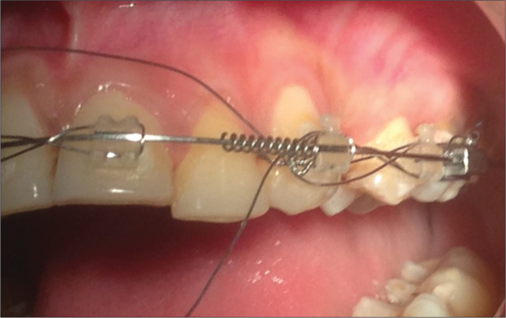
Figure 1
The ligature wire is placed without the placement of the arch wire
The arch wire is then placed with the open coil spring and the canine bracket is ligated with metal ligatures
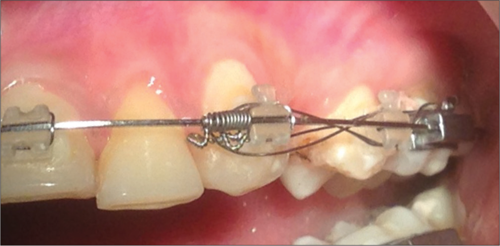
Figure 2
The arch wire is then placed with the open coil spring and the canine bracket is ligated with metal ligatures
The ends of the ligatures are brought forward and tied with the compression of the open coil spring
Figure 3
The ends of the ligatures are brought forward and tied with the compression of the open coil spring

This method is a modification to the original active laceback but with added advantages of Immediate reactivation of the laceback, Low force ratio and constant force application.

Financial support and sponsorship

Nil.

Conflicts of interest

There are no conflicts of interest.

References

  1. , , . Systemized Orthodontic Treatment Mechanics. Edinburgh: Mosby; .

Fulltext Views
7,952

PDF downloads
4,915
View/Download PDF
Download Citations
BibTeX
RIS
Show Sections