

Translate this page into:
S-template
Address for correspondence: Dr. Jitendra Bhagchandani, Department of Orthodontics and Dentofacial Orthopedics, Sardar Patel Post Graduate Institute of Dental and Medical Sciences, Lucknow, Uttar Pradesh, India. E-mail: jitendrabhagchandani@rocketmail.com
This article was originally published by Wolters Kluwer and was migrated to Scientific Scholar after the change of Publisher.
Abstract
Recently introduced Yen angle and W angle suggest the use of center of premaxilla (M) and mandibular symphysis (G) as landmarks for assessing sagittal jaw base discrepancies. These landmarks have been considered to be more stable than traditionally used points A and B. This article is an attempt to develop a simple tool that will enable the clinician to identify the centers of premaxilla and mandibular symphysis and hence the name Simplified template, i.e., S-template. This tool would aid the orthodontist in locating the centroids of maxilla and mandibular symphysis more easily and accurately than prefabricated templates.
Keywords
Template
point G
point M

INTRODUCTION
M point is defined as center of the premaxilla, while G point is the center of the largest circle that is tangent to the internal inferior, anterior and posterior surfaces of the mandibular symphysis.[1,2] Most of the recent cephalometric studies recognize the center of premaxilla (M point) and mandibular symphysis (G point)[3] to be stable landmarks against which the sagittal jaw discrepancy of the jaw bases should be assessed. These studies have made use of transparent template with circles to trace the center of the jaw bases. This clinical innovation referred to as simplified template, i.e., S-template; is an easy tool to make circles of varying diameters on the Powerpoint (version 2007, Microsoft Office) with their diameters increasing by 0.1 inch.
MATERIALS AND METHODOLOGY
A computer loaded with Microsoft Powerpoint Software version 2007
An inkjet printer. (HP Deskjet Ink Advantage 2060 K110)
An A4 size paper sheet
A pencil and a metal scale.
Methodology
Open the Microsoft PowerPoint software (version 2007)
Select a slide with a white background
Click on the insert bar and select shapes [Figure 1]
 Figure 1
- Click on the insert bar and select shapes
Insert a circle on the white template. Fill the circle with a color so that it’s easily reflected through the cellulose acetate tracing sheet [Figure 2]
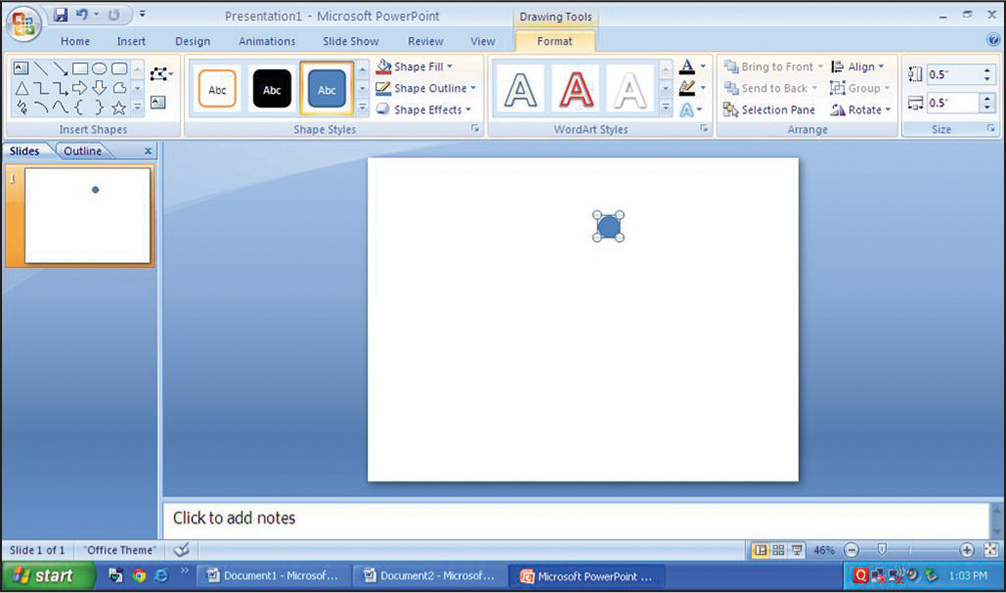 Figure 2
- Insert a circle on the white template and fill it with a color
Copy and paste such 10 circles on the slide
Adjust the dimensions of the circle by selecting the format bar and clicking on the extreme left side to designate the vertical and horizontal dimensions starting from 0.3 inch to 1.2 inch. Each circle increases in diameter by 0.1 inch [Figure 3]
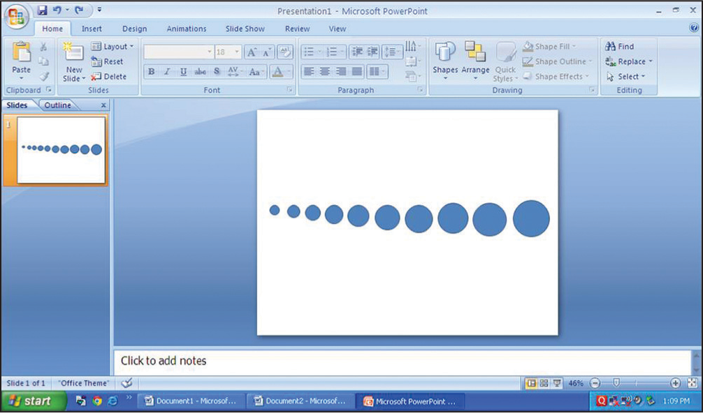 Figure 3
- Circles with increasing diameter by 0.1 inch
Take a print and mark the center of each circle with a fine black felt tipped pen [Figure 4]
 Figure 4
- Marking the center of the circles
The template is now prepared to be used against the cellulose acetate sheet to identify the center of pre maxilla and mandibular symphysis [Figure 5].
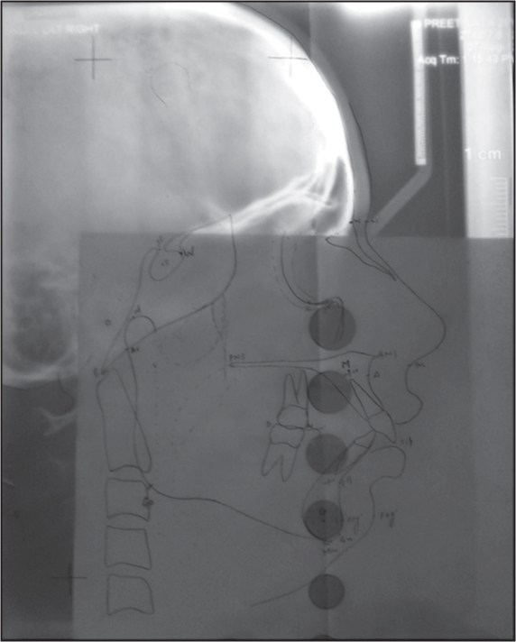 Figure 5
- Superimposing the cellulose acetate sheet over the template to identify point M and point G
CONCLUSION
This innovation could prove to be a helpful aid in measuring the angular parameters such as Yen angle and W angle. S-Template is easy to fabricate and more accurate.
Source of Support:
Nil.
Conflict of Interest:
None declared.


