Translate this page into:
Virtual planning for vertical control using temporary anchorage devices
Address for correspondence: Prof. Mauricio Accorsi, 526 Candido de Abreu Ave, #903B Curitiba, 80530-000 Parana, Brazil. E-mail: accorsi23@hotmail.com, accorsi@usp.br
This article was originally published by Wolters Kluwer and was migrated to Scientific Scholar after the change of Publisher.
Abstract
The new and innovative technologies are unprecedentedly improving the level of proficiency in orthodontics in the recent history of this area of expertise. The proliferation of advances, such as self-ligating systems, temporary anchorage devices, shape-memory wires, robotically wire bending, intraoral scanners, cone-beam computed tomography, bring the virtual planning, and confection of dental devices through CAD/CAM systems to the real world. In order to get efficiency and efficacy in orthodontics with these new technologies, we must understand the importance of systemically managed clinical information, medical, and dentistry history of the patients, including the images resources, which ensures the use of a communication that is assisted by the technology, with an interdisciplinary team so that the database is able to help and support the process of therapeutic decision-making. This paper presents the clinical case of a borderline patient for orthognathic surgery who had his final treatment planning supported by these new tools for three-dimensional diagnosis and virtual planning.
Keywords
Cone-beam computed tomography
decision-making process in orthodontics
three-dimensional imaging
INTRODUCTION
Cone-beam computed tomography (CBCT) has become popular in dentistry for virtual implant planning because of its objective and efficacy, since the paper published by Mozzo et al., in 1998,[1] describing the first cone-beam scanner dedicated to the maxillofacial area, the NewTom 9000 (QR s.l.r. Verona, Italy). After that, many other machines were released in the Dentistry market, but the most popular one is still i-CAT (Imaging Sciences, Hatfield, USA). At the same time, a number of software was also developed for applications in other areas such as Periodontics, Endodontics, Dental Sleep Medicine, Surgery, and Orthodontics. In addition, new protocols and analysis have been developed around the world for creating new standards and reference values to the craniofacial evaluation. These new technologies including the digital models and CAD/CAM systems are playing the role of game-changer in the entire diagnosis and therapeutic decision-making process, especially in Orthodontics. This paper presents the clinical case of a borderline patient for orthognathic surgery who had his final treatment planning supported by these new tools for three-dimensional diagnostic and virtual planning. Mini-screws were virtually positioned before the beginning of the treatment in order to support the vertical control approach. Furthermore, the airway volumetric analysis, the tongue posture, the TMJ, and the facial profile were taken in consideration for final decision. The paternalist approach in Orthodontics is losing support in the scientific environment, and the patient autonomy is being taken into consideration for the decision-making process, particularly in this case. The patient did not accept any treatment that would involve permanent teeth extraction or major surgery.
TREATMENT OBJECTIVES AND ALTERNATIVES
The patient was a 17-year-old male, and as shown in the initial records [Figure 1], the patient’s problem list presented a vertical facial pattern, poor facial profile with mandibular deficiency, anterior open bite supported by the bad tongue posture, flared upper and lower incisors, altered occlusal plane, and initial smile showing the inverted smile arc. The patient’s chief complaint was the poor esthetics and the difficulty to cut and chew food because of his anterior open bite. The medical and oral history was all right, and he had good oral health. After referring him to the diagnostic center, where he was submitted to a CBCT acquisition for the entire face (i-CAT with a 0.3 mm voxel size), three-dimensional reconstructions were performed for evaluating patient’s actual anatomy [Figure 2]. Airway trajectory was within the acceptable volume and minimal area, and the patient did not report any sleep problems or respiratory alterations.
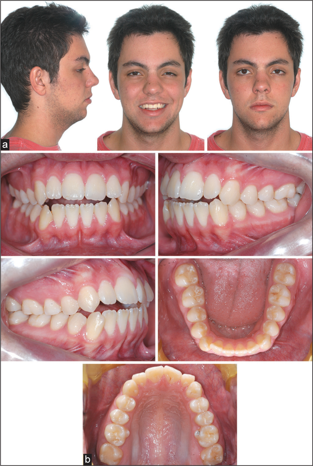
- Initial records: (a) Extra-oral pictures, (b) intra-oral pictures
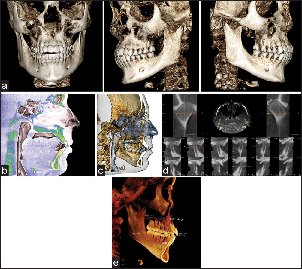
- Three-dimensional reconstructions and cross-sections obtained with Invivo5 (Anatomage Inc. San Jose, CA): (a) Dentoalveolar morphology; (b) tongue posture in sagittal clipping; (c) airway view and skeletal morphology in sagittal clipping. See the incisors projection; (d) TMJ in cross-sections (sagittal view); (e) sagittal clipping showing the upper and lower incisors angle related to maxillary and mandibular plane
The first treatment alternative presented to the patient was four first bicuspid extraction in order to correct the anterior open bite and the anterior torque and proclination. Considerations over the unpredictable effect in the facial profile were made, including the necessity of orthognathic surgery after the space closure and facial re-evaluation. The patient and his parents did not accept any of these alternatives; therefore, the second approach presented to them was related to vertical control and arch form modeling by means of a self-ligating system and posterior intrusion anchored by four mini-screws. This treatment alternative also included the patient’s referral to the speech therapist for tongue posture improvement and oral musculature adaptation. The temporary anchorage devices position was defined after the virtual planning by using the Invivo5 (Anatomage Inc. San Jose, USA) [Figure 3].
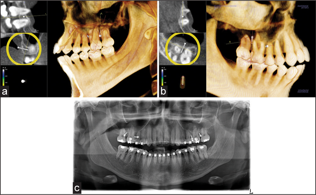
- (a) Virtual mini-screw planning obtained with Invivo5 (Anatomage Inc. San Jose, CA) internal view; (b) labial view; (c) panoramic radiography showing the temporary anchorage devices after placement
TREATMENT PROGRESS
After the initial leveling and alignment through round copper NiTi wires, and the Damon System, “Q” metallic and clear in the upper anterior (Ormco, Orange, USA), four mini-screws (Neodent, Curitiba, Brazil) were placed between the upper molar roots, mesiolabial and distolingual, and elastic chains were placed over the first molar crowns in order to activate the intrusion. [Figure 4]. The normal archwire sequence was performed for the case while the patient was taking his oral myotherapy exercises.
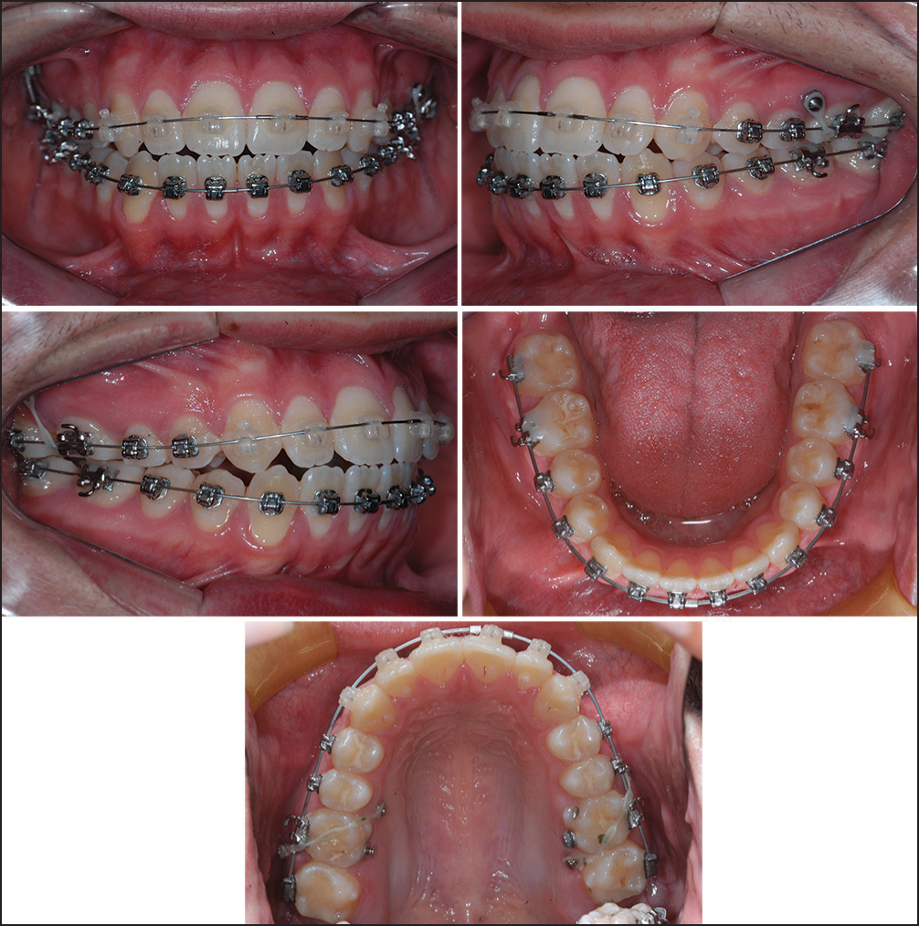
- Introral photograph after initial levelling and aligning
TREATMENT RESULTS
As shown in the final records [Figure 5], the anterior open bite no longer exists, and it is possible to note the improvement in the occlusal plane position and in the tongue posture. Note the images showing the treatment result taken by DICOM superimposition (Invivo5). Furthermore, it is possible to see the profile improvement and the smile arc in harmony with the lower lip [Figure 6].
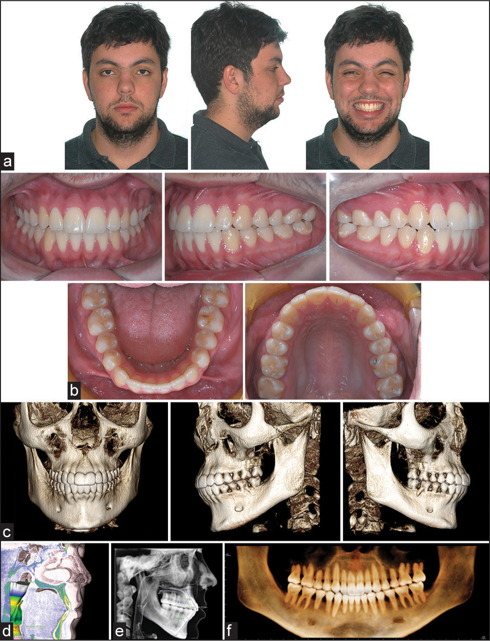
- Treatment outcome: (a) Extra-oral pictures; (b) intra-oral pictures; (c) dentoalveolar morphology; (d) tongue posture in sagittal clipping; (e) lateral cephalogram obtained with Invivo5 (Anatomage Inc. San Jose, CA) (f) coronal panoramic view obtained with Invivo5 (Anatomage Inc. San Jose, CA)
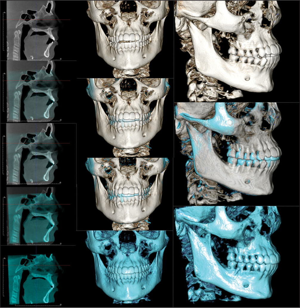
- Superimposition obtained with Invivo5 (Anatomage Inc. San Jose, CA)
DISCUSSION
The decision-making process has been changed in Orthodontics since the appearance of new and innovative technologies such as the CBCT, three-dimensional digital models, and the CAD/CAM systems that allows more precision in diagnosis and better treatment predictability. Furthermore, we are experiencing a paradigm shift in orthodontics by leaving the so-called “Angle’s paradigm”[2] behind and starting the “Oral Health Related Quality of Life” age.[3,4] The CBCT opens our eyes to very important findings that might be lost through conventional radiology exams. With the CBCT, we can see the dentition that is under formation, the facial bones structure, the dental alveolar morphology, the TMJs, the paranasal sinuses, the nasal cavity, the airways, and all other spatial interrelation among facial structures through three-dimensional virtual reality. We can also use the CBCT to make an early diagnosis of pathologies. This CBCT universe allows us to better understand the impact that our ability to make clinical correlations and to communicate with our patients and colleagues has, through a detailed definition of risks, benefits, and alternate treatment to our clients. Thus, the orthodontist assumes the responsibility of coordinating the interdisciplinary actions and the therapeutic decision-taken procedure throughout a variety of possible treatments that are available.
CONCLUSION
If we can reinforce the importance of paying attention and being careful with our health, the result will represent a great potential to improve our proficiency and our patients’ quality of life. And this reinforcement brings in itself a whole universe of new opportunities and possibilities, but it represents our big responsibility as experts to look for the required information and innovation in a reasonable and committed way. In this particular case, the patient demonstrated great satisfaction with the experience and especially with the results achieved.
Financial support and sponsorship
Nil.
Conflicts of interest
There are no conflicts of interest.
References
- A new volumetric CT machine for dental imaging based on the cone-beam technique: Preliminary results. Eur Radiol. 1998;8:1558-64.
- [Google Scholar]
- Enhancement Orthodontics: Theory and Practice. Ames, Iowa, USA: Wiley-Blackwell; 2007. p. :160.
- Diagnóstico 3D Em Ortodontia. A Tomografia Cone-Beam Aplicada. Nova Odessa: Editora Napoleão; 2001. p. :364.


