Translate this page into:
Treatment of severe maxillary cleft hypoplasia in a case with missing premaxilla with anterior maxillary distraction using tooth-borne hyrax appliance
Address for correspondence: Dr. Akshai Shetty, Department of Orthodontics and Dentofacial Orthopedics, R.V. Dental College and Hospital, Bengaluru, Karnataka, India. E-mail: akshaishetty@gmail.com
This article was originally published by Wolters Kluwer and was migrated to Scientific Scholar after the change of Publisher.
Abstract
Cleft orthodontics generally poses a challenge and a missing premaxilla adds to the difficulty in managing them. The lack of bone support and anterior teeth in a case with missing premaxilla accounts not only for difficulty in rehabilitation but also in increasing the maxillary hypoplasia. This article presents a case report where planned orthodontic and surgical management using distraction has helped treat a severe maxillary hypoplasia in a patient with missing premaxilla. The treatment plan and method can be used to treat severe maxillary hypoplasia and yield reasonably acceptable results for such patients.
Keywords
Anterior maxillary distraction
cleft maxillary hypoplasia
missing premaxilla
INTRODUCTION
Cleft maxillary hypoplasia is a common stigmata for cleft lip and palate repair. The severity of this deformity in bilateral cleft lip repair is more and its prevalence even higher.[1] The protruding premaxilla has been dealt in various ways with the evolution of bilateral cleft repair.[2] Unfortunately, some surgeons have also chosen sacrificing the premaxilla surgically during the definitive lip repair, as one of the treatment options in their pursuit to achieve a good lip repair. This leads to anterior anodontia and severe maxillary retrusion. This case report discusses one such rare case with a missing premaxilla and the treatment offered to the patient.
CASE REPORT
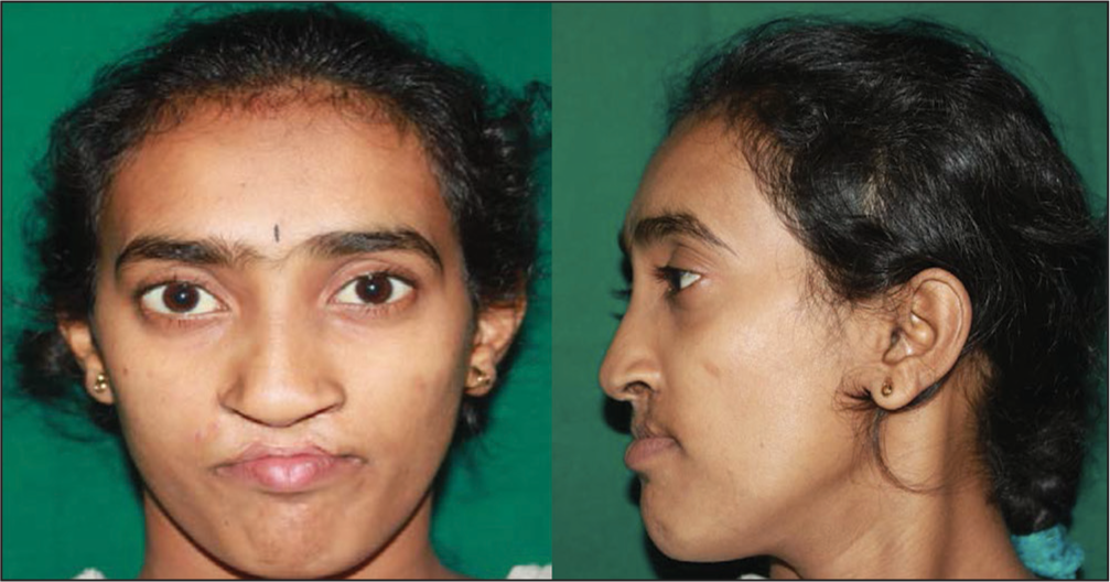
A 23-year-old female patient reported to our unit with a chief complaint of the backwardly placed upper jaw and difficulty in chewing. History revealed that she had bilateral cleft lip and palate for which she was operated in a rural setup. The cleft lip repair procedure was done at 6 months of age, and the premaxilla, unfortunately, was surgically removed to achieve closure of the lateral lip elements. On extra-oral examination, the patient had a concave profile due to the severe maxillary retrusion, and malar deficiency. The absence of columella widened ala base and flattened nasal tip contributed further to the dished-in face appearance [Figure 1].

- Pretreatment frontal and profile pictures
On intra-oral examination, a constricted maxilla with a cross-bite in the posterior region was noted. The anterior teeth (right central and lateral, left central lateral incisor and canine) were missing owing to the surgical removal of the premaxilla. The grossly decayed upper left second premolar was extracted, and the lower arch had missing first molars in both right and left the side that had led to mesial tipping of the second molars. The reverse jet had a discrepancy 12 mm [Figure 2].

- Cephalometric assessment of maxillary mandibular relationship
The speech of the patient was also a concern. The nasal air emission was prominent and intelligibility fair.
A composite cephalometric analysis was carried out which revealed that the size of the mandible was normal, thereby deriving the fault was in the maxilla. Since the premaxilla was surgically removed, we were unable to determine the maxillary cephalometric measurements with regard to point A. A horizontal growth pattern was also observed.
Joint consultation with the maxillofacial and orthodontic team helped us sketch the following objectives taking into consideration the clinical aspects and the diagnostic aids.
Correction of the mid-face deficiency and to achieve a positive overjet.
Leveling and aligning of teeth.
Upper arch expansion.
Replace missing teeth.
Achieve an ideal overbite.
The pre-surgical orthodontics comprised of initial leveling and aligning of both the upper and lower dental arches.
The exact assessment of maxillary advancement could not be determined due to the missing anteriors. Hence it was decided to place a temporary acrylic bridge using the 13 and 24 as lateral incisors abutments. The central incisor pontics were then placed to assess the actual discrepancy. Once the orthodontic goal was satisfactorily achieved, the patient was then prepared for the surgical maxillary advancement.
The technique used for the advancement was the brainchild of Dr. Gunasheelan Rajan. Although some cases have been published using this concept, but no one had reported a case where the premaxilla was missing.
A hyrax appliance was fabricated in the anterior-posterior relationship, that is, the axis of conventional hyrax used for the transverse maxillary expansion was inverted for distraction in anterior-posterior axis. The bands of the appliance were placed on the molars and the premolars on either side. The fit of the appliance was checked preoperatively [Figure 3].

- Temporary acrylic bridge for assessment of reverse overjet
Surgically, an anterior maxillary osteotomy posterior to the premolars on either side was done under general anesthesia. The prefabricated hyrax was then inserted and cemented on the operating table. The appliance was activated a few millimeters to check the ease of movement and the totality of the osteotomy cuts.
A period of consolidation of 5 days postoperatively was followed by daily activation of the appliance. The activation is 0.5 mm twice a day. The complete rotation of the hyrax screw adds up to 1 mm. This was carried out for 12 days and an additional 2 days (2 mm) for over-correction. A mild open bite developed during the course of distraction and box elastics were used anteriorly to correct it.
After a retention phase of 9 months with fixed orthodontic appliance the patient was debonded and referred to the prosthodontist to replace the temporary acrylic bridge anteriorly and also to rehabilitate the teeth in the extra space achieved postdistraction [Figure 4].

- Placement of hyrax for anteroposterior distraction
Two implants were placed in the first quadrant. The original existing premolar was shaped to form the canine and the space of distraction between the premolar (now canine), and the molar was used to place two implants replacing the first and second premolars.
In the second quadrant, the distraction space was used to place one implant and substitute as another premolar. The anterior teeth were now replaced with a ceramic fused to metal 4-unit bridge to form the anterior teeth (right and left lateral and central incisors) using the reshaped canine as lateral incisor abutments on the right side and reshaped first premolar on the left. The final rehabilitation of the patient was harmonic and esthetically acceptable [Figure 5]. The cephalometric analysis of pre- and post-operative radiographs showed improved maxillary, mandibular relationship with a gain of positive over jet [Figures 6 and 7]. The assessment of speech post-operatively by the speech therapist showed no change in hyper-nasality but an improved intelligibility and articulation.

- Space obtained postdistraction

- (a) Posttreatment extra-oral pictures. (b) Posttreatment intra-oral pictures

- Preoperative lateral cephalogram and postoperative lateral cephalogram
DISCUSSION
The growth disturbance related to cleft lip+/cleft palate repair has been well documented.[3] Maxillary hypoplasia both due to inherent growth disturbance along with surgical scarring is responsible for the poor maxillary mandibular relationship.[4,5]
Maxillary hypoplasia thus is the most common secondary problem to be dealt in cleft lip and palate patients.
Orthognathic surgery has been the mainstay in treating such deformities.[6] This was until distraction was tried as the treatment protocol.[7] The advantages of distraction over conventional orthognathic are many but specifically in cleft palate operated patients it helps in two ways. (1) Avoidance of relapse due to scar tissue.[8] (2) Preventing any further speech disturbance due to the increase in velopharyngeal insufficiency caused by sudden maxillary advancement.[9]
Distraction osteogenesis is a surgical technique that uses body’s own repair mechanisms for optimal reconstruction of the tissues. This would thus mean distraction should be an ideal treatment alternative to all maxillary hypoplastic cases but was not frequently opted for, due to the increased cost factor associated with internal maxillary distractors and low patient compliance.[10]
The problems of the internal maxillary distractor in cleft hypoplasia were satisfactorily dealt by the technical innovation of using the hyrax appliance in the anterior-posterior direction for advancing the maxilla.[11] This technique is easy and has the promise of correcting significant amount of reverse overjet (maxillary retrusion) using principles of distraction. The anterior maxilla after an anterior maxillary osteotomy can be distracted up to 18 mm with stable outcomes.[12] The maximum movement from a hyrax is 15 mm and for the additional movement we can remove the appliance re-wind the screw to zero and refabricate for further movement.
The obvious advantage in distracting only the anterior maxilla is keeping the posterior segment untouched.
In our case report, we dealt with a maxillary retrusion of 12 mm and ended up having a stable result. The challenge in our case was not just the maxillary hypoplasia but a missing premaxilla along with the associated anterior anodontia.
Bilateral cleft lip repair has a myriad of treatment protocols and many surgeons in the past believed in sacrificing the protruding premaxilla in their pursuit of getting a definitive lip closure.[2] The sacrificed premaxilla and the missing anterior ensured that the technical ease of anterior maxillary distraction is challenged in our case.
Rehabilitating the anterior teeth with acrylic temporary so as to gauge the actual anteroposterior discrepancy and the amount of correction required, helped us sketch the treatment plan. The anterior maxillary distraction followed by availability of bone in the segment of new bone formation between the molar and the premolar on either side not only helped in correcting the reverse overjet but also helped in increasing the arch length and space availability to restore the missing teeth.
Prosthodontic intervention for implants and anterior 4-unit bridge helped the patient get esthetic rehabilitation and a stable occlusion.
The speech of a patient is an important aspect while considering any advancement procedure of the maxilla. Conventional orthognathic surgeries increase the velopharyngeal space by stretching the palatal tissue.[13] In distraction, there is the gradual growth of the palatal tissue (histogenesis) that allows enough time for adaptation. In anterior maxillary distraction the distraction being between the molar and the premolar teeth has very little effect on the palatal tissue. In our case, the hyper-nasality of the patient postoperatively did not change, and the improvement in intelligibility and articulation can be attributed to improved maxillary mandibular relationship and increased maxillary arch width.
CONCLUSION
This case highlights the salient points of anterior maxillary distraction like-ease of operation, minimal speech disturbance, availability of new bone in maxillary arch for teeth rehabilitation, increase in maxillary arch length, improved maxillary mandibular relationship for a severe maxillary retrusion and stability of result.
Cleft care has been always described as an interdisciplinary treatment. The importance of joint consultation and treatment execution has been well rewarded by the result achieved in our case, and thus emphasizing that no single intervention can be completely gratifying.
Financial support and sponsorship
Nil.
Conflicts of interest
There are no conflicts of interest.
References
- Long-term effects of premaxillary setback on facial skeletal profile in complete bilateral cleft lip and palate. Cleft Palate J. 1985;22:97-105.
- [Google Scholar]
- Management of the premaxilla in bilateral clefts. J Oral Maxillofac Surg. 1983;41:518-24.
- [Google Scholar]
- A long-term retrospective outcome assessment of facial growth, secondary surgical need, and maxillary lateral incisor status in a surgical-orthodontic protocol for complete clefts. Plast Reconstr Surg. 2003;111:1-13.
- [Google Scholar]
- Factors that affect variability in impairment of maxillary growth in patients with cleft lip and palate treated using the same surgical protocol. J Plast Surg Hand Surg. 2011;45:188-93.
- [Google Scholar]
- Maxillary sequelae in cleft patients. Causes of maxillary hypoplasia and possible prevention. Rev Stomatol Chir Maxillofac. 2007;108:297-300.
- [Google Scholar]
- Frequency of Le Fort I osteotomy after repaired cleft lip and palate or cleft palate. Cleft Palate Craniofac J. 2007;44:396-401.
- [Google Scholar]
- A meta-analysis of cleft maxillary osteotomy and distraction osteogenesis. Int J Oral Maxillofac Surg. 2006;35:14-24.
- [Google Scholar]
- Factors related to relapse after Le Fort I maxillary advancement osteotomy in patients with cleft lip and palate. Cleft Palate Craniofac J. 2001;38:1-10.
- [Google Scholar]
- Velopharyngeal changes after maxillary advancement in cleft patients with distraction osteogenesis using a rigid external distraction device: A 1-year cephalometric follow-up. J Craniofac Surg. 1999;10:312-20.
- [Google Scholar]
- Clinical controversies in oral and maxillofacial surgery: Part one. Maxillary distraction osteogenesis for advancement in cleft patients, internal devices. J Oral Maxillofac Surg. 2008;66:2592-7.
- [Google Scholar]
- Anterior maxillary distraction by tooth-borne palatal distractor. J Oral Maxillofac Surg. 2007;65:1044-9.
- [Google Scholar]
- Anterior maxillary distraction using a tooth-borne device for hypoplastic cleft maxillas – A pilot study. J Oral Maxillofac Surg. 2011;69:e542-8.
- [Google Scholar]
- Predictors of velopharyngeal insufficiency after Le Fort I maxillary advancement in patients with cleft palate. J Oral Maxillofac Surg. 2011;69:2226-32.
- [Google Scholar]


