

Translate this page into:
Evaluating condylar position in different skeletal malocclusion patterns: A cephalometric study
Address for Correspondence: Dr. Sandesh S. Hegde, 254, Kumar Hostel, S.D.M College of Dental Sciences, Sattur, Dharwad - 580 009, Karnataka, India. E-mail: shegde19.sh@gmail.com
This article was originally published by Wolters Kluwer and was migrated to Scientific Scholar after the change of Publisher.
Abstract
Context
The cranial base and variations in its morphology affect the anterior-posterior positioning of jaws causing changes in the glenoid fossa and condylar position.
Aims
To evaluate the condylar position in patients with different skeletal sagittal malocclusion patterns.
Materials and Methods
Pretreatment lateral cephalometric radiographs of 112 subjects (both males and females) were categorized into three classes (Class I, Class II, Class III) based on their ANB angulation and studied for N-S-Ar (saddle angle), S-Ar-Go (articular angle), S-Ar (posterior cranial base length).
Statistical Analysis
Shapiro-Wilk test was done to check for normality of the distribution of values. Groups were evaluated using parametric tests (one-way ANOVA). Significance for all tests was predetermined as P < 0.05.
Results
N-S-Ar and S-Ar-Go and also S-Ar did not vary significantly in all the three classes. N-S-Ar and S-Ar-Go angles have shown a significant negative correlation in all the three classes.
Conclusions
There is no significant difference in condylar position in different skeletal malocclusion patterns. N-S-Ar and S-Ar-Go angles show a negative correlation in any skeletal malocclusion pattern.
Keywords
Cephalometric
condylar position
skeletal malocclusion

INTRODUCTION
The cranial base and the variations in its morphology have always been assumed to affect the anterior-posterior positioning of jaws. To study the relationship between cranial base morphology and malocclusion has been of keen interest to researchers, some of the early studies were done by Huxley,[1] Young,[2] Renfroe[3] and Moss.[4] The existence of a significant relationship between cranial base morphology and jaw relationship was demonstrated first by Bjork[5] using cephalometric radiographs and in later times by Melsen.[6] The maxilla and mandible articulate with different parts of the cranial base, hence variations in growth and orientation of the cranial base region leads to differential movement of the maxilla and mandible causing changes in glenoid fossa and condylar position. Droel and Isaacson,[7] Baccetti et al.[8] have analyzed the position of the glenoid fossa in subjects with differential sagittal and vertical features, but very limited data is available on the significance of condylar position in different sagittal malocclusion patterns. This study aims at evaluating condylar position in patients with different skeletal sagittal malocclusion patterns.
MATERIALS AND METHODS
Pretreatment lateral cephalometric radiographs of 112 subjects (both males and females) were selected. Each of the lateral cephalograms were studied and categorized into three classes (Class I, Class II, Class III) based on their ANB angulation.
Skeletal sagittal relationships on the basis of the ANB values:
(Class I = 2° < ANB <4°; Class II = ANB >4°; Class III = ANB <2°) Ballard.[9]
Three groups were formed; Class I (37), Class II (52), Class III (23). All the subjects were normodivergent and belonged to the age group between 15 and 30 years. Patients with craniofacial growth disorders were excluded from the study.
CEPHALOMETRIC ANALYSIS
The lateral cephalometric radiographs of each subject were taken with (KODAK 9000C extraoral imaging). All subjects were positioned in the cephalostat with the sagittal plane at a right angle to the path of the X-rays, the Frankfort plane parallel to the horizontal, the teeth in centric occlusion, and the lips slightly closed. The radiographs were hand-traced and measured by the same investigator. All tracings and measurements were repeated in 1-weeks’ time to check for any error.
The following landmarks were used for cephalometric analysis:
Point A (A): It is the deepest point on the midline between the anterior nasal spine and alveolar crest between the two central incisors.
Point B (B): It is the deepest point between the alveolar crest of the mandible and the mental process.
Articulare (Ar): Intersection of the posterior border of the condyle and the posterior cranial base.
Sella (S): Centre of sella turcica.
Nasion (N): Most anterior point on frontonasal suture.
Gonion (Go): Lowermost point at the intersection of mandibular and ramal planes.
The following measurements were used:
Angular measurements for the assessment of sagittal growth pattern
SNA, SNB, ANB.
Angular measurements for the assessment of cranial base flexure:
N-S-Ar (Saddle angle), S-Ar-Go (Articular angle).
Linear measurements for the assessment of position of condyle:
S-Ar (Posterior cranial base length).
Statistical analysis
The mean and standard deviations were estimated for each cephalometric variable in each group [Figures 1-3]. The majority of the cephalometric variables were normally distributed according to Shapiro-Wilk test [Table 1] hence the differences between the groups were evaluated using parametric tests (One-way ANOVA). Significance for all tests was predetermined as P < 0.05. All statistical analysis was performed using Statistical Package for Social Sciences software package (SPSS Base 10.0 for Windows User’s Guide. SPSS Inc., Chicago, IL, USA).

- Saddle angle (degree) values for three malocclusion classes
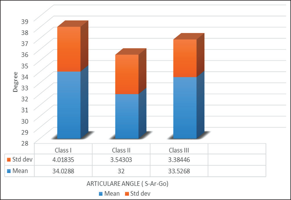
- Articular angle (degree) values for three malocclusion classes
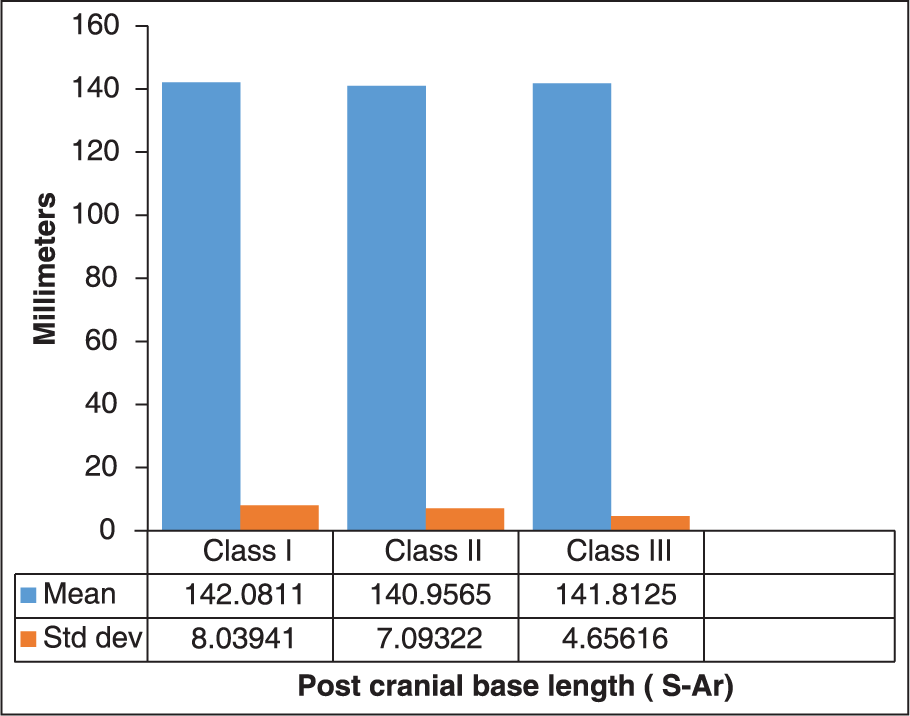
- Posterior Cranial Base length (millimetres) for three malocclusion classes
| Class | Shapiro-Wilk | ||
|---|---|---|---|
| Statistic | df | Sig. | |
| N-S-Ar | |||
| Class 1 | .976 | 37 | .609 |
| Class 2 | .979 | 52 | .467 |
| Class 3 | .907 | 23 | .036 |
| S-Ar-Go | |||
| Class 1 | .920 | 37 | .011 |
| Class 2 | .986 | 52 | .789 |
| Class 3 | .848 | 23 | .002 |
| Post cr l | |||
| Class 1 | .982 | 37 | .787 |
| Class 2 | .981 | 52 | .554 |
| Class 3 | .952 | 23 | .324 |
RESULTS
The following results were deduced:
Saddle angle (N-S-Ar) and Articular angle (S-Ar-Go) and also Posterior Cranial base length (S-Ar) did not vary significantly in all the three classes [Table 2].
Table 2 One-way ANOVA to test the signifi cance difference between different classesCephalometric parameter Mean square F Sig. N-S-Ar Between groups 3.157 .154 .858 Within groups 20.546 Total S-Ar-Go Between groups 10.675 .217 .806 Within groups 49.263 Total Post cr l Between groups 34.458 2.549 .083 Within groups 13.518 Total Since P values are all greater than 0.05 the classes do not diff er in all the three measurement
N-S-Ar and S-Ar-Go angles have shown a significant correlation in all the three classes [Graph 1]. There exists a negative correlation between the two angles and the regression equation deduced is:
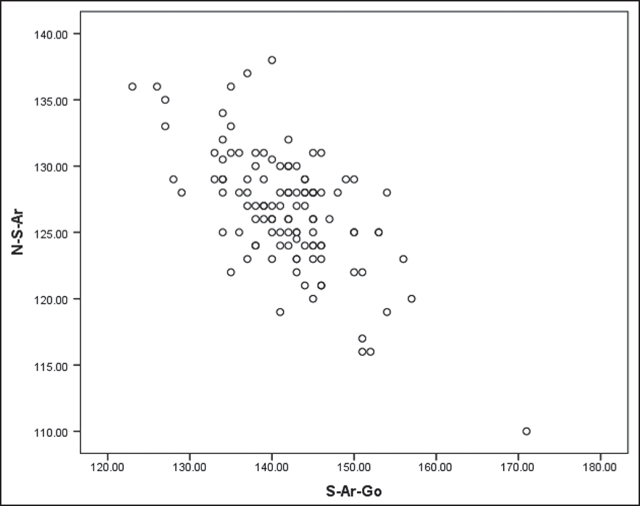 Graph 1
- Graph shows negative correlation between S-Ar-Go and N-S-Ar
DISCUSSION
The cause of orthodontic problems arising from antero-posterior malrelationship of jaws has been mainly attributed to changes in its size form and position (Hopkin et al.).[10] Scott[11] has stated three main factors that influence facial prognathism: Opening of the cranial base angle, the relative forward movement of components like maxilla and mandible to the cranium, and the amount of surface deposition along the facial profile between nasion and menton. Changes in cranial base morphology has been put forth as a possible indicator of skeletal malocclusion by several researchers who have found a significant relationship in between cranial base and antero-posterior jaw position (Anderson and Popovitch,[12] Wihelm et al.,[13] Bacon et al.,[14] Dibbets et al.[15] and Singh et al.[16]). Enlow[17] has shown that growth of maxilla is under the influence of the cranial base while the mandible acts in a more independent way, although its articulation at the glenoid fossa does provide potential for influence from the cranial base; hence variations in the cranial base morphology may cause changes in the position of glenoid fossa and the condyle. Numerous studies have been carried out to check the correlation between cranial base flexure and skeletal malocclusion, but with contradicting conclusions, studies by Bjork,[5] Hopkin et al.,[10] Dibbets et al.,[15] Bacon et al.[14] and Järvinen[18] have proved a significant relationship between the two while studies by Klocke and Nanda,[19] Polat and Kaya,[20] Lewis and Roche,[21] Kasai et al.[22] and Dhopatkar et al.[23] have proved otherwise. In a study done by Kerr and Adams[24] it has been suggested that cranial base flexure influences mandibular prognathism by determining the anteroposterior position of the condyle relative to the facial profile. Baccetti et al.[8] concluded that glenoid fossa is associated with a more posterior position in class II when compared to class III skeletal malrelation. No such studies have been carried out to determine changes in condylar position.
In the study three measurements, two angular (N-S-Ar and S-Ar-Go) and one linear measurement (S-Ar) were used as parameters to determine condylar position in Class I, Class II and Class III malocclusion patterns using lateral cephalograms. N-S-Ar (saddle angle) which is also known as the cranial base flexure angle helps determine the changes in the cranial base angulations. Posterior cranial base (S-Ar) was used to determine the distance of the condyle from the S. In both cases Ar (articular) was used as the posterior limit instead of the Ba (basion) point as it marks the intersection of the condyle and the posterior cranial base. There always has been disagreement whether the posterior base should be measured from Ba or Ar. Bjork[5] and Hopkin[10] have both advocated the use of Ar, rather than basion, because of its ease of identification. Varjanne and Koski[25] have discouraged the use of Ar because of its remoteness from the cranial base and suggested basion as the more appropriate choice. Similarly, Kerr and Adams[24] used basion to measure the cranial base angle. Bhatia and Leighton[26] who have published figures for N-S-Ba and N-S-Art angles as well as the S-Ba and S-Ar distances found the growth patterns as described by use of basion or Ar to be similar. Seward[27] has also explained the use of Ar point over basion point as a parameter for determining condylar position. Point Ar is displaced backward and downward during growth and it is affected by the direction of condylar growth and of mandibular rotation (Björk;[28] Popovich and Thompson,[29] Björk and Skieller).[30]
No significant difference was seen in any of the three cephalometric variables in all the three classes (Cl I, Cl II and Cl III) of sagittal malrelations. It should be noted that the temporo-mandibular joint is positioned at the lateral edges of the cranial base and is, in fact, considerably separated spatially from the midsagittal plane on which cephalometric analyses are based. It is likely, therefore, that changes in the cranial base angle may not be directly translated to the mandibular articulation (Dhopatkar et al.).[23] The correlation analysis revealed a negative relationship between the N-S-Ar and S-Ar-Go angles in all the three classes.
Along with the parameters analyzed in the study it is important for us to consider other factors such as role of soft tissue a causative influence in development of different malocclusion patterns, Solow and Kreiborg[31] stated that factors inducing cranial extension, impairment of nasal airflow influence craniofacial development. DAttilio et al.[32] proved a statistically significant correlation with mandibular position and length, overjet, and the mandibular plane angle to the cervical curvature. Festa et al.[33] showed a significant correlation between distal jaw position, sagittal mandibular length, and increased cervical lordosis.
Hence, it can be concluded that the condylar position did not significantly change in different antero-posterior jaw malrelations and to further confirm the finding studies with larger number of subjects and better imaging techniques could be advocated.
CONCLUSION
There is no significant difference in condylar position in different skeletal malocclusion patterns.
N-S-Ar and S-Ar-Go angles have shown a significant negative correlation in all the three classes.
References
- Evidence As to Man’s Place in Nature. London: Williams and Norgate; 1863.
- A contribution to the study of the Scottish skull. Trans R Soc Edinb. 1916;51:347-453.
- [Google Scholar]
- A study of the facial patterns associated with class I, class II division 1, class II division 2 malocclusions. Angle Orthod. 1948;18:12-5.
- [Google Scholar]
- Correlation of cranial base angulation with cephalic malformations and growth disharmonies of dental interest. NY State Dent. 1955;24:452-4.
- [Google Scholar]
- The cranial base: the postnatal development of the cranial base studied historically on human autopsy material. Acta Odontologica Scandinavica. 1974;32(62):57-71.
- [Google Scholar]
- Some relationships between the glenoid fossa position and various skeletal discrepancies. American Journal of Orthodontics. 1972;61:64-78.
- [Google Scholar]
- Glenoid fossa position in different facial types; a cephalometric study. Br J Orthod. 1997;24:55-9.
- [Google Scholar]
- Some bases for aetiology and diagnosis in orthodontics. Dental Record. 1948;68:6-14.
- [Google Scholar]
- The cranial base as an aetiological factor in malocclusion. Angle Orthod. 1968;38:250-5.
- [Google Scholar]
- Dento-facial Development and Growth. Oxford: Pergamon Press; 1967.
- Relation of cranial base flexure to cranial form and mandibular position. Am J Phys Anthropol. 1983;61:181-7.
- [Google Scholar]
- A comparison of cranial base growth in Class I and Class II skeletal patterns. Am J Orthod Dentofacial Orthop. 2001;119:401-5.
- [Google Scholar]
- The cranial base in subjects with dental and skeletal Class II. Eur J Orthod. 1992;14:224-8.
- [Google Scholar]
- Finite element analysis of the cranial base in subjects with Class III malocclusion. Br J Orthod. 1997;24:103-12.
- [Google Scholar]
- Saddle angle and maxillary prognathism: A radiological analysis of the association between the NSAr and SNA angles. Br J Orthod. 1984;11:209-13.
- [Google Scholar]
- Role of cranial base flexure in developing sagittal jaw discrepancies. Am J Orthod Dentofacial Orthop. 2002;122:386-91.
- [Google Scholar]
- Changes in cranial base morphology in different malocclusions. Orthod Craniofac Res. 2007;10:216-21.
- [Google Scholar]
- Pubertal spurts in cranial base and mandible: Comparisons between individuals. Angle Orthod. 1985;55:17-30.
- [Google Scholar]
- Relationship between cranial base and maxillofacial morphology. Eur J Orthod. 1995;17:403-10.
- [Google Scholar]
- An investigation into the relationship between the cranial base angle and malocclusion. Angle Orthod. 2002;72:456-63.
- [Google Scholar]
- Cranial base, sagittal jaw relationship and occlusion. A radiological-craniometric appraisal. Proc Finn Dent Soc. 1982;78:179-83.
- [Google Scholar]
- A Manual of Facial Growth. Oxford: Oxford University Press; 1993.
- Normal and abnormal growth of the mandible. A sinthesis of longitudinal cephalometric implant studies over a period of 25 years. Eur J Orthod. 1983;5:1-46.
- [Google Scholar]
- Soft-tissue stretching: A possible control factor in craniofacial morphogenesis. Scand J Dent Res. 1977;85:505-7.
- [Google Scholar]
- Cervical lordosis angle measured on lateral cephalograms; findings in skeletal class II female subjects with and without TMD: A cross sectional study. Cranio. 2004;22:27-44.
- [Google Scholar]
- Relationship between cervical lordosis and facial morphology in Caucasian women with skeletal class II malocclusion: A cross sectional study. Cranio. 2003;21:121-9.
- [Google Scholar]


