Translate this page into:
Evaluation of natural head position changes in the treatment of class II malocclusions with functional appliance

*Corresponding author: Nazanin Nasr, Dental Student, Department of Orthodontics, Shahid Sadoughi University of Medical Sciences, Yazd, Iran. nsrnazanin2000@gmail.com
-
Received: ,
Accepted: ,
How to cite this article: Yassaei S, Vakil F, Nasr N. Evaluation of natural head position changes in the treatment of class II malocclusions with functional appliance. APOS Trends Orthod. 2024;14:170-7. doi: 10.25259/APOS_63_2023
Abstract
Objectives:
The position of the head to the neck is affected by various factors, including respiratory tract features, temporomandibular joint disorders, type of occlusion, and physical aspects. In addition, malocclusion can negatively affect the head-neck position. The present study aimed to evaluate changes in natural head position following treatment of Class II malocclusions with Farmand II functional appliance.
Material and Methods:
The present study was implemented as a historical cohort with the before and after design. Pre- and post-treatment lateral cephalometric radiographs of 33 skeletal Class II patients treated with the Farmand II functional appliance were included in the present study. The cephalometric analysis was done by tracing the related lines and landmarks. Facial angles and angles determining the natural head and neck position including craniohorizontal, craniocervical, craniovertical (CCV), and cervical curvature (CCI) were measured. Collected data were entered in Statistical Package for the Social Sciences (SPSS)-version 17 software. Paired t-test was used to evaluate the changes in cephalometric indices before and after treatment. P < 0.05 was defined to be statistically significant for all the tests.
Results:
The results of the present study, showed a significant reduction in the mean values of A point, nasion, B point (ANB) angle and angle of convexity following treatment (P < 0.001); also, Sella-nasion/Odontoid process tangent (SN/OPT) angle showed a substantial decrease in treated patients (P = 0.033), which is indicative of head flexion following treatment. Moreover, the decrease in the mean values of the craniocervical angles, including True vertical line (NL/VER) and Sella-nasion/true vertical line (SN/VER), suggests downward bending of the head in patients treated with this appliance (P-values of 0.029 and 0.012, respectively). The odontoid process tangent/cervical vertebra tangent (OPT/CVT) angle showed an insignificant increase (P = 0.260).
Conclusion:
The present study showed that patients treated with the Farmand II functional appliance tend to flex their heads and maintain their cervical vertebra in a more upright and straight position.
Keywords
Class II malocclusion
Micrognathisms
Cervical vertebrae
Removable orthodontic appliance
INTRODUCTION
Diagnosis and treatment planning in orthodontic patients require some collection of a comprehensive database. Radiographic evaluation of the dental and skeletal structures most commonly done using analyses of lateral cephalograms and panoramic views is part of the database collection process. In the analysis of lateral cephalograms, reference lines such as anterior cranial base Sella-Nasion (SN) line and Frankfort horizontal (FH) line are commonly used to evaluate the skeletal structures in different spatial planes. Despite their widespread use, these reference lines are subjected to specific limitations.[1,2]
Natural head position (NHP), is defined as a standard and repeatable position of a standing person, in which eyes are focused on a point at a distance at eye level. NHP is commonly used both in medical and dental clinical evaluation by plastic surgeons, as well as oral and maxillofacial surgeons and orthodontists.[3]
NHP shows the highest repeatability among adults and children, females and males, and Caucasians and non-Caucasians, with 4° of variance.[4] In 1998, Cooke and Wei reported the repeatability of NHP with 2° of variance.[5]
Studies have shown several factors affecting the head and neck relationship, including respiratory tract disorder, temporomandibular joint disorders, physical features age, sex, and facial morphology.[6]
The head and neck relationship can also be affected by the presence of malocclusion.[1,5-7] Hellsing et al. reported that the cervical angle in individuals who have well-aligned teeth is 3–5° greater than in those who have anterior crowding of 2 mm or more in the maxillary and mandibular arch.[8] In recent studies, including D’Attilio et al.,[9] Hedayati et al.,[10] and Yassaei et al.,[11] it was shown that in Class II malocclusion patients, the head is tilted backward (extension), and skeletal Class III patients present a forward rotation of the head relative to the spine (flexion).
Furthermore, different studies have shown that the position of cervical vertebrae can change following orthodontic surgery treatment using.[12-17]
Cho et al.[12] and Tejaswi et al.[13] reported that patients with Class III malocclusion who have undergone Lefort 1 surgery and bilateral sagittal split osteotomy (BSSO) setback had changes in their NHP, which tended toward a head extension. Phillips et al. reported that patients with Class III malocclusion who have undergone Lefort 1 surgery and BSSO setback tended toward head flexion.[14]
Lin and Edwards evaluating the effect of mandibular advancement in skeletal Class II malocclusion due to the deficient mandible on the head position reported that surgical intervention using BSSO leads to forward movement of the cervical vertebrae and the NHP will be achieved in a more upright position.[15] Furthermore, Achilleos et al.[16] and Schendel and Epker[17] reported that following mandibular advancement in the surgical approach, there would be a forward movement of the cervical vertebrae that leads to cervical lordosis and head flexion.
Samlliene et al. evaluated the effect of treatment with twin block appliances on body posture in Class II malocclusion subjects, reporting the posture body changes during treatment with the functional appliance were an expression of physiologic growth, not a response to functional therapy.[18]
On the other hand, recent studies[19-22] have evaluated the NHP in patients with skeletal Class II malocclusion before and after the treatment using different functional appliances (twin block, activator, and Frankel). Farmand is a kind of functional appliance that is like the bionator appliance. It was designed and introduced in 1972 by Farmand S.M. and registered at Loyola University.[23,24] This appliance is used by some of Iran’s orthodontists. Therefore, the present study aimed to evaluate the NHP in patients with skeletal Class II malocclusion before and after the treatment using Farmad II functional appliance.
MATERIAL AND METHODS
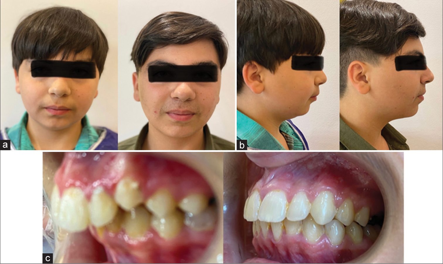
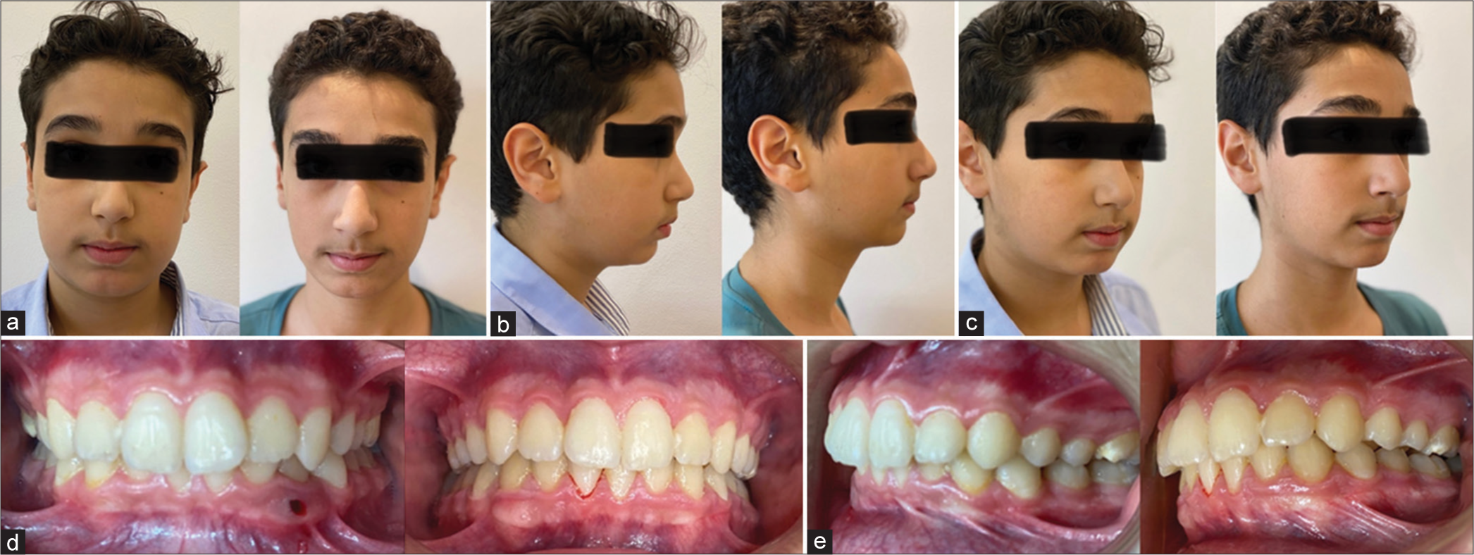
The present study was implemented as a pre-post historical cohort study. It was approved by the Ethical Review Board with the code of IR.SSU.REC.1400.020. Lateral cephalometric radiographs of thirty-three skeletal class II patients with an average age of 11.2 ± 1.3 years were selected (13 boys of 11-14 years old, and 20 girls of 10-13 years old). All patients were treated with palatal expansion for 3 months and then Fa-II appliance for 11–13 months. Pre- and post-treatment photographs are shown in [Figures 1-3].

- Pre- and post-treatment photographs: (a) frontal view, (b) profile view, (c) oblique view, (d) smile view, and (e) intraoral view.

- Pre- and post-treatment photographs: (a) frontal view, (b) profile view, and (c) lateral view intraoral.

- Pre- and post-treatment photographs: (a) frontal view, (b) profile view, (c) oblique view, (d) intraoral view, and (e) lateral view of intraoral.
Cephalograms were taken in the standard position (maximal intercuspation of teeth, lips in light contact, and NHP). Bilateral ear rods were inserted into the external auditory meatus to stabilize the head during exposure. The patients were instructed to stand in NHP and to stare at their own eyes reflected in a 60 × 90 cm mirror at a distance of 1 m. All cephalograms were taken by the same operator. The cephalometric evaluation was done by tracing the related lines and landmarks.
Inclusion criteria
The following criteria were included in the study:
Overjet ≥5 mm before treatment
Canines and molars in Class II relationships
A point, nasion, B point (ANB) angle >4.5°
Mandibular deficiency: Body length < Se-N +3 mm and/or saddle angle >128°
Skeletal Class II malocclusion due to the deficient mandible
Horizontal or normal growth pattern
Treated with the Farmand II functional appliance [Figure 4a and b]
Lateral cephalometry provided in NHP
Accessibility of lateral cephalometry before and after the treatment in patients’ files
No previous history of orthodontic treatment
No syndromic or medically compromised patient
Achieving normal overjet (2–3 mm) after treatment.

- (a) Farmand II functional appliance, (b) patient treated with Farmand II functional appliance
Exclusion criteria
The following criteria were excluded from the study:
Not obvious cervical vertebrae
Low-quality lateral cephalometry
Patients with facial asymmetry
Patients with a history of mouth breathing and tonsillar hyperplasia.
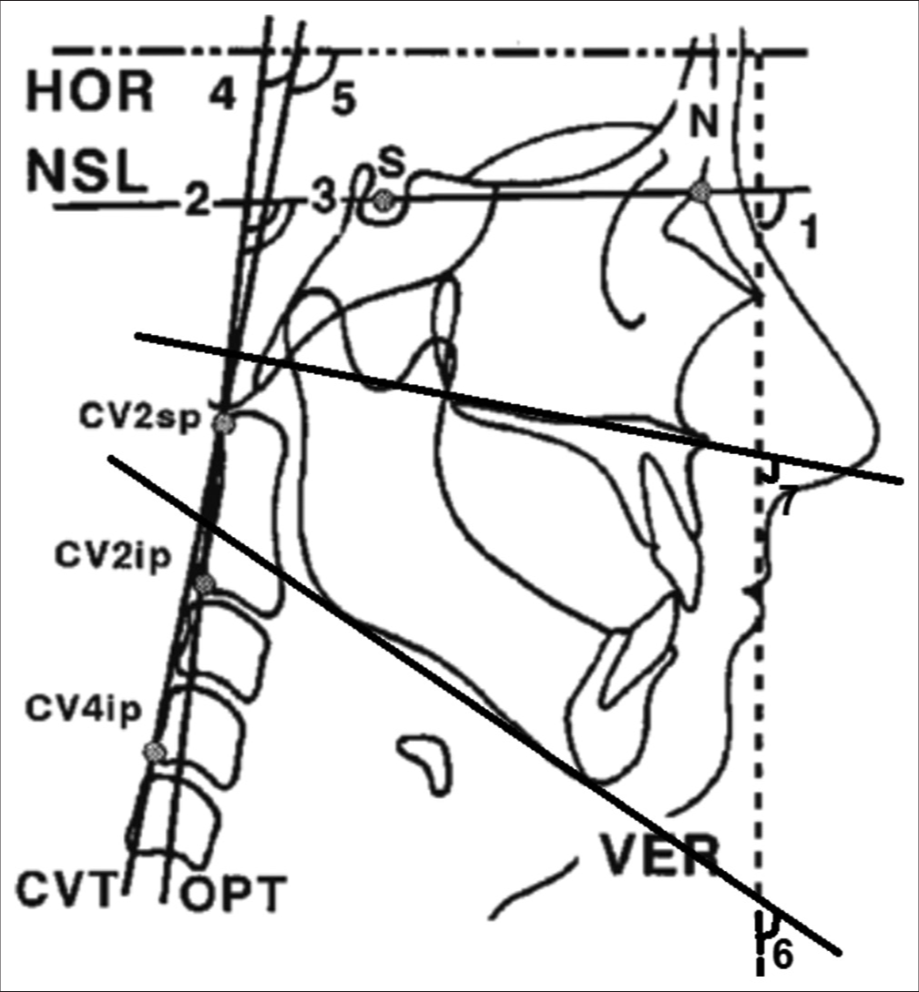
Thirteen reference points, including nine points on the skull and four points on the spinal vertebrae, were marked on the tracing paper using a pointed-tip pen. The shade of the sagged chain in the lateral cephalometry was considered the true vertical plane, and the true horizontal plane was defined by drawing a perpendicular line to the true vertical plane. In addition, odontoid process tangent (OPT), cervical vertebra tangent (CVT), palatal, occlusal, SN, and mandibular planes were drawn, and intended angles, including craniohorizontal (Odontoid process tangent/True horizontal line [OPT/HOR] and Cervical vertebra tangent/True horizontal line [CVT/HOR]), craniovertical (CCV) (GO-GN/true vertical line; True vertical line [NL/VER]; and Sella-nasion/true vertical line [SN/VER]), craniocervical (Sella-nasion/Odontoid process tangent [SN/OPT], Sella-nasion/Cervical vertebra tangent [SN/CVT], ANS-PNS/odontoid process tangent, and NL/Cervical vertebra tangent/Odontoid process tangent/[CVT/OPT]), and cervical curvature (CCI) (CVT/OPT), were measured by an instructed researcher [Table 1 and Figure 5]. All measurements were done manually and re-confirmed by the assessor 1 week after the preliminary calculation, and the mean values of the angles were considered.
| CV2sp: The most superior and posterior point on the odontoid process of cervical vertebrae CV2ip: The most inferior and posterior point on the corpus of the second cervical vertebrae CV4ip: The most inferior and posterior point on the corpus of the fourth cervical vertebrae Anterior cranial base: SN (SNL): The anterior-posterior extent of the anterior cranial base Occlusal plane: The line interlinked from the contact point of the distal cusps of the maxillary and mandibular first molars to the contact point of the cusps of the maxillary and mandibular first premolars ANS-PNS (NL) (Palatal Plane): The line connecting the most posterior point of the hard palate to the most anterior point of the anterior nasal spine GO-GN (ML) (Mandibular Plane): Includes the inferior border of the mandible, tangent to the inferior mandibular border from the Me OPT: The line interlinking CV2sp and CV2ip points CVT: The line interlinking CV2sp and CV4ip points True vertical line: An external reference line, which is usually defined as the shade of the sagged chain in the lateral cephalometry True horizontal line: An extracranial reference line is defined by drawing a perpendicular line to the true vertical line Craniovertical angles: including SN/VER, NL/VER, and ML/VER angles Craniohorizontal angles: including OPT/HOR and CVT/HOR angles Craniocervical angles: including SN/OPT, SN/CVT, NL/OPT, and NL/CVT angles Cervical curvature: The angle between OPT and CVT |
SNL: Sella-Nasion line, ANS-PNS: Anterior nasal spine- Posterior nasal spine, GO-GN: Gonion-Gnathion, CV2sp: Second cervical vertebra superior point, CV2ip: Second cervical vertebra inferior point, CV24ip: 4thcervical vertebra inferior point, OPT: Odontoid process tangent, CVT: Cervical vertebra tangent, SN/VER: Sella -nasion/true vertical line, NL/VER: ANS-PNS/True vertical line, ML/VER: GO-GN/True vertical line, OPT/HOR: Odontoid process tangent/True horizontal line, CVT/HOR: Cervical vertebra tangent/True horizontal line, SN/OPT: Sella-nasion/Odontoid process tangent, SN/CVT: Sella-nasion/Cervical vertebra tangent, NL/OPT: ANS-PNS/Odontoid process tangent, NL/CVT: ANS-PNS/Cervical vertebra tangent.

- A sample tracing of angles determining the natural head and neck position. HOR: Horizontal, CVT: Cervical vertebra tangent, OPT: Odontoid process tangent, VER: Vertical, CV2sp-Second cervical vertebra superior point, CV2ip: Second cervical vertebra inferior point, CV24ip: 4th cervical vertebra inferior point, SNL: Sella Nasion line, N: Nasion, S: Sella, (1) SNL/VER angle, (2) SNL/OPT angle, (3) SNL/CVT angle, (4) OPT/HOR angle, (5) CVT/HOR angle, (6) ML/VER angle, (7) NL/VER angle.
Statistical analysis
The sample size was calculated according to a 95% confidence level and the power of the test is 90%, with standard deviation before the intervention S1 = 3.12 and after the intervention S2 = 13.6 (for ML). Hence, 36 samples were needed.[23]
Mean values and standard deviation were calculated for each parameter. Data analysis was performed using IBM Statistical Package for the Social Sciences (SPSS) Statistics Version 17.0 software. Paired t-tests were used to evaluate changes in cephalometric indices before and after treatment. P < 0.05 was defined to be statistically significant for all the tests.
RESULTS
In the present study, lateral cephalometric radiographs of 33 patients with skeletal Class II malocclusion, before and after the treatment done by Farmand II functional appliance, were evaluated. The correlation coefficients of the studied angles were positive and significant in all cases (P < 0.05), which means that there was a positive correlation between the angles in all planes before and after treatment.
Our findings showed that the mean value of ANB angles before and after treatment was 9.97 and 3.91°, respectively, showing a significant reduction in this angle during treatment [Table 2]. Moreover, we found a significant decrease in the mean value of the angle of convexity following treatment (P < 0.001).
| Angles | Number of samples | Before treatment | After treatment | Changes | P-value | |||
|---|---|---|---|---|---|---|---|---|
| Mean | SD | Mean | SD | Mean | SD | |||
| ANB | 33 | 9.97 | 1.59 | 3.91 | 1.53 | 5.06 | 1.27 | <0.001 |
| Angle of convexity | 33 | 9.72 | 3.43 | 7.52 | 3.04 | 2.21 | 2.34 | <0.001 |
ANB: A point- Nasion- B point, SD: Standard deviation
There was a slight reduction in the mean value of ML/VER after the treatment that was not significant (71 and 70.24°, respectively, before and after treatment). Furthermore, there was a significant reduction in the mean values for true vertical line (NL/VER) angle before and after treatment (P = 0.029); similarly, 2.09° reduction in the mean values of SN/VER angle before and after treatment was considered significant [Table 3].
| Craniovertical angles | Number of samples | Before treatment | After treatment | Changes | P-value | |||
|---|---|---|---|---|---|---|---|---|
| Mean | SD | Mean | SD | Mean | SD | |||
| ML/VER | 33 | 71.00 | 5.36 | 70.24 | 5.86 | 0.76 | 4.6 | 0.351 |
| NL/VER | 33 | 93.24 | 4.43 | 91.18 | 5.14 | 2.06 | 5.17 | 0.029 |
| SN/VER | 33 | 101.1 | 5.35 | 99.09 | 5.07 | 2.09 | 4.49 | 0.012 |
SN/VER: Sella -nasion/True vertical line; NL/VER: ANS-PNS/True vertical line; ML/VER: GO-GN/True vertical line, SD: Standard deviation
The results also showed that the application of Farmand II functional appliance in skeletal Class II patients led to a slight reduction in the mean values of NL/OPT, NL/CVT, and SN/CVT angles, none of which were significant (P > 0.05). However, SN/OPT angles showed a reduction, from 101.42 before treatment to 98.76° after treatment, which was considered significant [Table 4].
| Craniocervical angles | Number of samples | Before treatment | After treatment | Changes | P-value | |||
|---|---|---|---|---|---|---|---|---|
| Mean | SD | Mean | SD | Mean | SD | |||
| NL/OPT | 33 | 93.33 | 8.76 | 90.94 | 9.29 | 2.39 | 7.85 | 0.090 |
| NL/CVT | 33 | 98.97 | 8.22 | 96.55 | 7.56 | 2.42 | 7.45 | 0.071 |
| SN/CVT | 33 | 106.5 | 10.06 | 104.7 | 8.85 | 1.85 | 7.37 | 0.160 |
| SN/OPT | 33 | 101.42 | 9.86 | 98.76 | 9.29 | 2.67 | 6.87 | 0.033 |
NL/OPT: ANS-PNS/Odontoid process tangent, NL/CVT: ANS-PNS/Cervical vertebra tangent, SN/CVT: Sella-nasion/Cervical vertebra tangent, SN/OPT: Sella-nasion/Odontoid process tangent, SD: Standard deviation.
Regarding craniohorizontal angles, an insignificant increase was demonstrated in both OPT/HOR and CVT/HOR angles comparing before and after treatment values (P > 0.05) [Table 5].
| Craniohorizontal angles | Number of samples | Before treatment | After treatment | Changes | P-value | |||
|---|---|---|---|---|---|---|---|---|
| Mean | SD | Mean | SD | Mean | SD | |||
| OPT/HOR | 33 | 90.18 | 8.67 | 91.09 | 8.74 | -0.91 | 7.92 | 0.515 |
| CVT/HOR | 33 | 85.06 | 8.84 | 85.67 | 8.62 | -0.61 | 7.66 | 0.653 |
OPT/HOR: Odontoid process tangent/True horizontal line, CVT/HOR: Cervical vertebra tangent/True horizontal line, SD: Standard deviation.
Finally, considering CCI, the results showed an increase in the mean values of odontoid process tangent/cervical vertebra tangent (OPT/CVT) angle before and after treatment. However, this difference was not considered statistically significant (P > 0.05) [Table 6].
| Cranio curvature angles | Number of samples | Before treatment | After treatment | Changes | P-value | |||
|---|---|---|---|---|---|---|---|---|
| Mean | SD | Mean | SD | Mean | SD | |||
| OPT/CVT | 33 | 5.77 | 2.57 | 6.27 | 2.21 | -0.5 | 2.51 | 0.260 |
Paired t-test, OPT/CVT: Odontoid process tangent/Cervical vertebra tangent, SD: Standard deviation.
DISCUSSION
The results of the present study showed that the mean value for ANB angle, which is representative of changes in the maxillomandibular skeletal relationship during treatment had a 5.06° reduction indicating the impact of the Farmand II functional appliance on improving the form of the face during treatment. Moreover, the mean value of angle of convexity showed 2.21° reduction that is significant and shows improvement in profile and skeletal relationship during treatment. These findings were consistent with the results of a study by Yassaei and Soroush[23] and Yassaei et al.[24]
The results also showed an insignificant decrease in the mean values of craniocervical angles, which indicate the relationship of the horizontal line of the head (SN and NL) to the spine (CVT and OPT) (P = 0.09). However, a study with a larger sample size was needed for a more definitive conclusion. These results show that treatment with the Farmand II functional appliance flexes the head downward. In addition, SN/OPT angle showed a significant decrease following the application of this appliance, which offers head flexion during treatment. Moreover, a significant reduction in the CCV angles indicates that patients with skeletal Class II malocclusion bend their heads downward following treatment with the Farmand II functional appliance. Finally, OPT/CVT angle showed an insignificant increase, which offers a more straight and upright cervical vertebrae position in these patients following the application of the Farmand II functional appliance.
Ohnmeiß et al.[19] reported no significant change in the angle between the spinal plane (OPT), and the horizontal lines of the head (Cranial base, palatal plane, and mandibular plane) following treatment with activator and bite jump functional appliances. The results of our study are similar to theirs.
Aglarci[20] evaluated the position of the head and cervical vertebrae during treatment with twin block. Similar to the previous studies, they showed no alterations in the angle between the upper section of the spine and horizontal lines of the head following treatment of patients using Class II functional twin block, so the results are consistent with the previous studies.
In a study by Kamal and Fida,[21] a cephalometric evaluation of the position of the head and spine during functional treatment with a twin block appliance was done, and a significant increase in the SN/OPT angle was reported, which can be considered a probable indicator of the upright position of the upper section of the spine. Furthermore, Tecco et al.[22] showed a significant increase in SN/OPT and SN/CVT angles following functional treatment using the Frankel appliance. This shows head extension on the upper cervical vertebrae and upward rotation of the head. These results controvert the results of our study.
CCI angles (OPT/CVT) showing the curvature of the cervical vertebrae demonstrated a 0.5° increase, which was not considered statistically considerable. However, in Aglarci’s study,[20] contradictory results were achieved, and they indicate a significant increase in this proportion that shows a change in the position of the middle cervical vertebrae. On the other hand, studies evaluating the effect of the twin block, and the Frankel appliances on this index supported the results of our research and showed no significant change.[21,22]
Lin and Edwards,[15] evaluating the effect of mandibular advancement in skeletal Class II malocclusion due to the deficient mandible on the head position, reported that surgical intervention using BSSO leads to forward movement of the cervical vertebrae increase in Sella-Nasion-C2 (SNC2) and alteration in the head position due to the increased craniocervical inclination. This indicated that following mandibular advancement, forward movement of the head helps in maintaining balance. At the same time, the muscular structure of the neck tilts the cervical vertebrae to a more forward position. Therefore, the NHP will be achieved in a more upright position.
Achilleos et al.[16] reported that following mandibular advancement in the surgical approach, there would be a forward movement of the cervical vertebrae that leads to cervical lordosis and head flexion. Hence, the results of our study are alike the studies which are about the impact of surgical advancement of the mandible on the head and neck position by Schendel and Epker,[17] Lin and Edwards,[15] and Achilleos et al.[16]
CONCLUSION
Regarding the results of the present study, it can be inferred that patients with Class II skeletal malocclusion tend to bend their head downward (flexion), and maintain their cervical vertebrae in a more upright and straight position following treatment with the Farmand II functional appliance.
Ethical approval
The authors declare that they have taken the Institutional Review Board approval and the approval number is IRB-2022/68-39.
Declaration of patient consent
The authors certify that they have obtained all appropriate patient consent.
Conflicts of interest
There are no conflicts of interest.
Use of artificial intelligence (AI)-assisted technology for manuscript preparation
The authors confirm that there was no use of artificial intelligence (AI)-assisted technology for assisting in the writing or editing of the manuscript and no images were manipulated using AI.
Financial support and sponsorship
Nil.
References
- Cephalometric features of class III malocclusion. Rev Med Chir Soc Med Nat Iasi. 2015;119:1153-60.
- [Google Scholar]
- Five-year reproducibility of natural head posture: A longitudinal study. Am J Orthod Dentofacial Orthop. 1990;97:489-94.
- [CrossRef] [PubMed] [Google Scholar]
- Fifteen-year reproducibility of natural head posture: A longitudinal study. Am J Orthod Dentofacial Orthop. 1999;116:82-5.
- [CrossRef] [PubMed] [Google Scholar]
- Effect of bilateral sagittal split ramus osteotomy setback on the soft palate and pharyngeal airway space. Int J Oral Maxillofac Surg. 2008;37:419-23.
- [CrossRef] [PubMed] [Google Scholar]
- The reproducibility of natural head posture: A methodological study. Am J Orthod Dentofacial Orthop. 1988;93:280-8.
- [CrossRef] [PubMed] [Google Scholar]
- Craniocervical morphology and posture in Australian aboriginals. Am J Phys Anthropol. 1982;59:33-45.
- [CrossRef] [PubMed] [Google Scholar]
- Morphology of the first cervical vertebra in children with enlarged adenoids. Eur J Orthod. 1985;7:93-6.
- [CrossRef] [PubMed] [Google Scholar]
- Cervical and lumbar lordosis and thoracic kyphosis in 8, 11 and 15-year-old children. Eur J Orthod. 1987;9:129-38.
- [CrossRef] [PubMed] [Google Scholar]
- Evaluation of cervical posture of children in skeletal class I, II, and III. Cranio. 2005;23:219-28.
- [CrossRef] [PubMed] [Google Scholar]
- Comparison of natural head position in different anteroposterior malocclusions. J Dent (Tehran). 2013;10:210-20.
- [Google Scholar]
- Cephalometric association of mandibular size/length to the natural head position. Int J Med Invest. 2019;8:51-62.
- [Google Scholar]
- Changes in natural head position after orthognathic surgery in skeletal Class III patients. Am J Orthod Dentofacial Orthop. 2015;147:747-54.
- [CrossRef] [PubMed] [Google Scholar]
- To evaluate the changes in Natural Head Position after orthognathic surgeries in class II patients and class III patients. Int J Disaster Risk Reduct. 2019;2:1-9.
- [Google Scholar]
- The effect of orthognathic surgery on head posture. Eur J Orthod. 1991;13:397-403.
- [CrossRef] [PubMed] [Google Scholar]
- Changes in natural head position in response to mandibular advancement. Br J Oral Maxillofac Surg. 2017;55:471-5.
- [CrossRef] [PubMed] [Google Scholar]
- Surgical mandibular advancement and changes in uvuloglossopharyngeal morphology and head posture: A short-and long-term cephalometric study in males. Eur J Orthod. 2000;22:367-81.
- [CrossRef] [PubMed] [Google Scholar]
- Results after mandibular advancement surgery: An analysis of 87 cases. J Oral Surg. 1980;38:265-82.
- [Google Scholar]
- Effect of treatment with twin-block appliances on body posture in class II malocclusion subjects: A prospective clinical study. Med Sci Monit. 2017;20:343-52.
- [CrossRef] [PubMed] [Google Scholar]
- Therapeutic effects of functional orthodontic appliances on cervical spine posture: A retrospective cephalometric study. Head Face Med. 2014;10:7.
- [CrossRef] [PubMed] [Google Scholar]
- Evaluation of cervical spine posture after functional therapy with twin-block appliances. J Orthod Res. 2016;4:8-12.
- [CrossRef] [Google Scholar]
- Evaluation of cervical spine posture after functional therapy with twin-block appliances: A retrospective cohort study. Am J Orthod Dentofacial Orthop. 2019;155:656-61.
- [CrossRef] [PubMed] [Google Scholar]
- Evaluation of cervical spine posture after functional therapy with FR-2: A longitudinal study. Cranio. 2005;23:53-66.
- [CrossRef] [PubMed] [Google Scholar]
- Changes in hyoid position following treatment of Class II division1 malocclusions with a functional appliance. J Clin Pediatr Dent. 2008;33:81-4.
- [CrossRef] [PubMed] [Google Scholar]
- Effects of Twin-Block and Faramand-LL appliances on soft tissue profile in the treatment of Class II division 1 malocclusion. Int J Orthod Milwaukee. 2014;25:57-62.
- [Google Scholar]


