Translate this page into:
New concept of physiologic anchorage control
Address for correspondence: Prof. Tian-Min Xu, Peking University School of Stomatology, China. E-mail: tmxuortho@163.com
This article was originally published by Wolters Kluwer and was migrated to Scientific Scholar after the change of Publisher.
Abstract
Molar anchorage loss in extraction case is believed due to the reaction of mechanical force applied to retract anterior teeth. While it may be close to truth in adult patients, it is certainly not true in adolescents. Studies on molar growth show upper molar move forward as mandible growing forward, probably through intercuspation force. Hence, for adolescents, molar anchorage loss shall consist of two parts. One is from retraction force — mechanical anchorage loss; another from biologic force — physiologic anchorage loss. Since physiologic anchorage loss is caused by the continuous biologic force, the strategy of physiologic anchorage control (PAC) is different from the strategy of mechanical anchorage control. A new PAC method is introduced in this article that can reduce the headgear and temporary anchorage device used as sagittal anchorage dramatically in orthodontic clinic.
Keywords
Anchorage loss
mandible growth
physiologic anchorage control
Anchorage is the footstone for moving malocclusion teeth in orthodontic treatment. To make this footstone stable, orthodontists designed different anchorage methods, such as stationary anchorage,[1] headgear, anchorage bends,[2] anchorage preparation,[3] cortical anchorage,[4] transpalatal arch (TPA),[5] Nance arch,[6] and implant anchorage[7-9] in the orthodontic history. The most striking star among the above anchorage measures is the implant anchorage. Before it appears, the maximum anchorage is defined as molar forward displacement less than one-fourth of a premolar extraction space in an orthodontic extraction case.[10,11] However, implant anchorage can make 0 mm molar anchorage loss theoretically. Orthodontists then have the strongest tool to challenge the alveolar limit, especially in cases with skeletal protrusion. The problem is: Is that good for periodontal health?
Figure 1 is an adult bimaxillary protrusion case. To reduce her protrusive lips, we extracted her four first premolars and retracted the anterior teeth with implant anchorage. After both the patient and doctor were satisfied with her profile, we stopped retraction, took impression and cone beam computed tomography (CBCT) as stage records. Superimposing digital study casts on unloaded mini-screw implants, we can see upper molars have not moved mesially at all, and upper incisors retracted apparently.
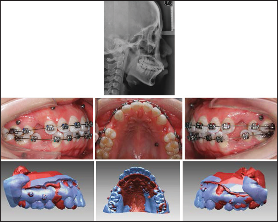
- Adult bimaxillary protrusion treated with four premolar extraction and miniscrew anchorage. Superimposition at the miniscrew interface sows retraction of incisors with no anchor loss
Since her upper molars did not move mesially at all, we can call this case an absolute anchorage case. Then we were wondering where will be the roots of incisors. Checking her CBCT image, we can see apparent root resorption of upper central incisors and alveolar defect on lingual side [Figure 2a]. We can also see alveolar fenestration of upper left lateral incisor apex on the labial side [Figure 2b]. If we check her lower incisor, more than half of the root on the lingual side is out of alveolar bone [Figure 2b]. More than 70 years ago, Dr. Tweed, considered labial limits, advocated extraction treatment.[12] Today, when we have absolute anchorage tools, should not we consider the lingual limits? In physiologic anchorage control (PAC), the fi rst principle is respecting the alveolar limits. Moreover, this limit should not be considered statically physical limit, but physiologic limit changing with growth and bone reconstruction.
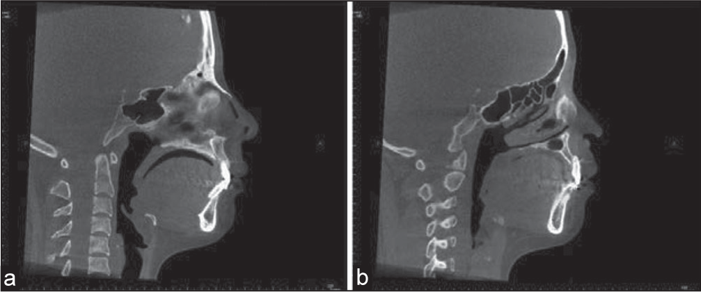
- (a) After retraction of incisors with miniscrew implant, alveolar bone of both upper and lower central incisors lost on lingual sides and upper central incisor root apex resorbed; (b) Alveolar fenestration of upper lateral incisor root
If we accept physiologic alveolar limits and believe the treatment objects, we, orthodontists, deal with are not static but dynamic, we will change our paradigm of anchorage control. Orthodontists used to attribute molar mesial displacement in extraction case to the reaction of mechanical force applied to retract anterior teeth. While it may be close to truth in adult patients, it is certainly not true in adolescents. Solow’s study[13] on 14 girls with Bjork metallic implant shows that upper molar moved 8 mm downward and 3 mm forward on average during 9-25 years. He also indicated for the forward mandibular rotation cases, upper molar can move forward much more than the average. One case in his sample, upper molar moved forward more than 7 mm, almost a premolar’s width. Johnston’s recent study[14] on 39 growing subjects from Bolton-Brush growth center shows upper molar move forward approximate to the amount of mandible outgrowing the maxilla. From his sample, upper molar moved forward about 2 mm (more than one-fourth of a premolar extraction space, the amount close to our maximum anchorage control definition) during 11-13 or 12-14, the common age periods at which we treat malocclusion. Moreover, our own prospective randomized clinical trial shows anchorage loss more for growing patient before the growth peak than after the peak.[15] We can, therefore, deduce as mandible outgrowing maxilla, it brings upper teeth moving forward through intercuspation force. Other biologic forces to move molar mesially include horizontal components of bite force and periodontal ligament force. Hence, we believe molar anchorage loss during orthodontic treatment shall actually consist of two parts, one part is from reaction, we call it mechanical anchorage loss; and another part is from growth or other biologic force, we call it physiologic anchorage loss. Then, the question is why we should differentiate physiologic anchorage loss and mechanical anchorage loss.
The way we perceive how molar anchorage loss affects the strategy that we adopt to control anchorage. If we believe anchorage loss is totally due to reaction, we deal with reaction only. We use TPA or Nance arch to disperse the reaction on molars; we use headgear to resist the reaction on molars; and we use implant anchorage to bypass the reaction. Take implant anchorage as an example, if we believe reaction is the only source of anchorage loss, detouring reaction from molar to miniscrew will theoretically keep molar stable. Figure 3a shows a high angle Class II adolescent, we extracted her four fi rst premolars and inserted miniscrew implants as anchorage. The fi rst wire is 0.014 nickel-titanium (NiTi), canines lacebacked to miniscrews to relieve anterior crowding. After 2 months, we took impression and made a digital cast superimposition on the stable area that we established by an implant marked study.[16] Although there was no anchorage burden on upper molars, superimposition showed upper molar tipped forward to loss anchorage. Why is that?
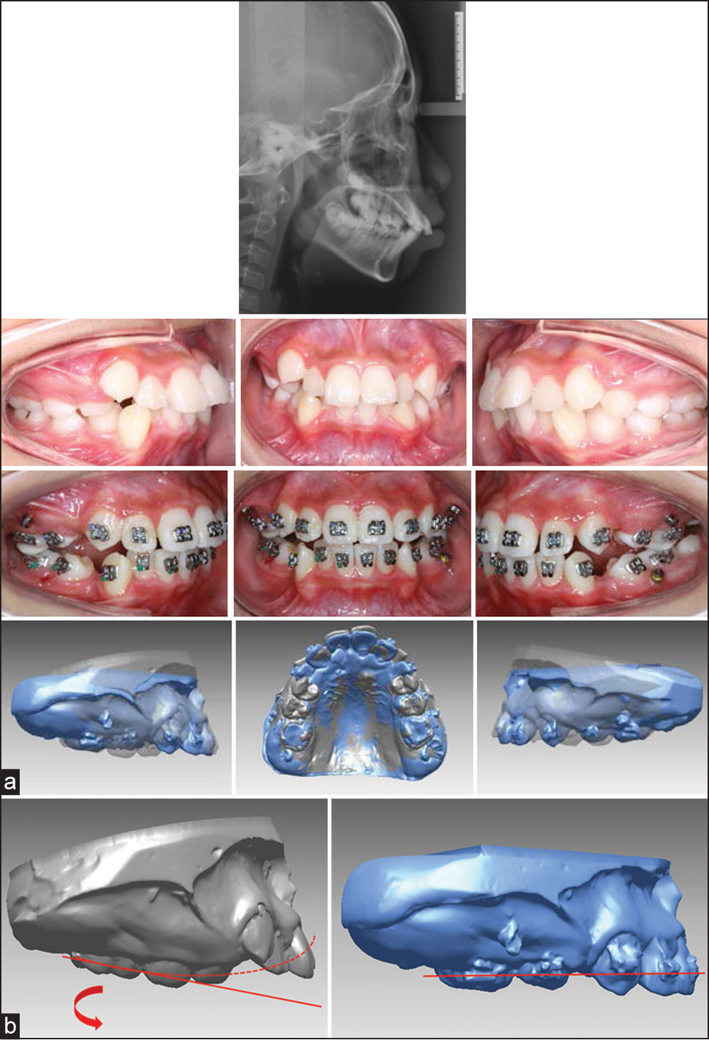
- (a) 12-year-old high angle Class II case. Upper molars tipped forward in 2 months even with mini-screw implants as anchorage. (b) Engagement of 0.014˝ nickel titanium wire resulted in the molar anchor loss
Let’s check our appliance fi rst. In modern straight wire appliance, we use 0° buccal tube. For patients with apparent curve of Spee in upper arch like this patient, engaging a straight archwire in anterior teeth will tip upper molars forward to lose anchorage [Figure 3b].
Why this happened so easy? Let’s look at how upper molar grow without appliance. Baumrind’s study[17] on implant superimposition shows upper molars tipped forward during growth [Figure 4]. Martinelli, et al. study[18] on Burlington Class II growth sample shows upper molar tipped 2.8° on average from 12-14 years. Moreover, our own maximum anchorage sample treated with headgear, upper molar tipped forward 7.2° on average during 2.5 years.[15] Hence, upper molar tipping forward is actually normal growth pattern, our modern straight wire appliance just accelerates this kind of physiologic anchorage loss. If we realize the existence of physiologic anchorage loss, we can then try to prevent it with new approaches.
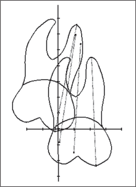
- Upper molar growth pattern traced using American metallic implant growth sample. The initial observation age is 8.5 and the last is 15.5; between them are 10.5 and 12.5, respectively. One unit of scale equals 2 mm. Horizontal frame of reference is Downs occlusal plane
Our own way to prevent physiologic anchorage loss is using a special cross buccal tube that consists of two tubes,[19] one −25° round tube and another −7° rectangular tube [Figure 5]. Two tubes cross in front, so we call it cross buccal tube (or brief it as XBT). It is a substitute of a tipback bend in traditional Tweed or Begg, the irreplaceable part is the gentle, continuous moment provided by the initial thin NiTi wire pairing with it. If the physiologic anchorage loss is caused by continuous biologic force, the preventive measures should certainly adopt the same force pattern. If we can prevent molar forward tipping with tipback tube during aligning stage and keep it with proper mechanics until we start to retract anterior teeth, molars will be in a relative tipback position, a posture similar to Tweed anchorage preparation that has been proved good for anchorage control by Tweed practitioner.
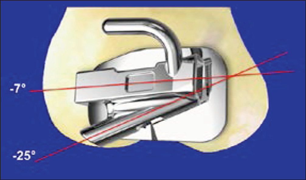
- The cross buccal tube designed to prevent physiologic anchorage loss
Figure 6 is an example treated with the PAC approach. The patient was a 13-year-old girl. She was Class II with lip protrusion [Figure 6a]. We extracted her three fi rst premolars and one second premolar on the lower left side because of the full Class II relationship on that side. Bonding XBT on molars and multi-level low friction brackets[20] on six anterior teeth, we used 0.014 NiTi as the initial arch wire [Figure 6b]. Forty days later, her anterior crowding was relieved and we then bonded her upper 5’ and 7’ and changed arch wire to 0.016 NiTi with the curve of Spee to keep upper molars in a relative backward tipping position [Figure 6c] while aligning upper dentition. Following the same procedure, we aligned her lower dentition and proceeded to the space closing stage [Figure 6d]. We fi nished the whole treatment in 20 months and improved her profile [Figure 6e]. Structure superimposition on maxilla shows her incisors retracted apparently and upper molar anchorage controlled very well, only a little forward tipping [Figure 6f]. Comparing to our headgear sample with 0° buccal tube, upper molar tipped 7.2° forward on average,[15] the XBT tube design serve our purpose to prevent physiologic anchorage loss and then enhanced total anchorage control fairly well.
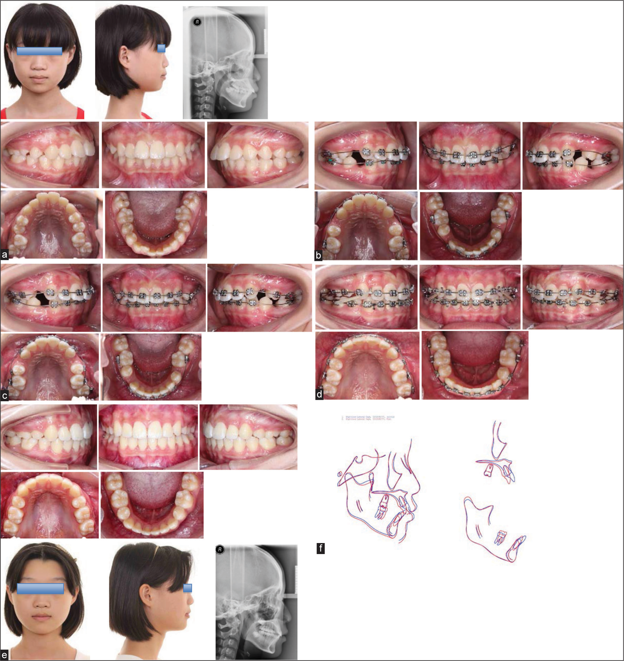
- A Class II protrusion case treated with PAC technique
PAC is a new concept, it has reduced headgear and temporary anchorage device used as sagittal anchorage dramatically in our clinic. I hope it can help more orthodontists in their clinic.
Parts of content quote from author’s lectures in 2012 and 2013 AAO Annual Session.
Financial support and sponsorship
Supported by Beijing Municipal Science & Technology Commission No. Z141107002514054.
Conflicts of interest
Dr. Xu is the inventor of the PAC system.
References
- Treatment of Malocclusion of the Teeth (7th ed). Philadelphia, PA: S.S. White Dental Manufacturing Co.; 1907.
- Creative wire bending — The force system from step and V bends. Am J Orthod Dentofacial Orthop. 1988;93:59-67.
- [Google Scholar]
- The application of the principles of the edgewise arch in the treatment of malocclusions: II. Angle Orthod. 1941;6:12-67.
- [Google Scholar]
- Effect of the transpalatal arch during extraction treatment. Am J Orthod Dentofacial Orthop. 2008;133:852-60.
- [Google Scholar]
- The limitations of orthodontic treatment: I. Mixed dentition diagnosis and treatment. Am J Orthod Oral Surg. 1947;33:177-223.
- [Google Scholar]
- Paradigm shifts in orthodontic treatment with mini-implant anchorage. APOS Trends Orthod. 2015;5:56.
- [Google Scholar]
- The use of miniscrew implants for temporary skeletal anchorage in orthodontics: A comprehensive review. Oral Surg Oral Med Oral Pathol Oral Radiol Endod. 2007;103:e6-15.
- [Google Scholar]
- Mini-implant anchorage for the orthodontic practitioner. Am J Orthod Dentofacial Orthop. 2008;133:621-7.
- [Google Scholar]
- Biomechanics and Esthetic Strategies in clinical orthodontics. St. Louis, MO: Elsevier Saunders; 2005. p. :196.
- Textbook of Orthodontics (1st ed). New Delhi: Jaypee; 2004. p. :264.
- Clinical Orthodontics. St. Louis: Mosby; 1966.
- Continued eruption of maxillary incisors and first molars in girls from 9 to 25 years, studied by the implant method. Eur J Orthod. 1996;18:245-56.
- [Google Scholar]
- Class II malocclusion: The aftermath of a “perfect storm”. Semin Orthod. 2014;20:59-73.
- [Google Scholar]
- Randomized clinical trial comparing control of maxillary anchorage with 2 retraction techniques. Am J Orthod Dentofacial Orthop. 2010;138:544.e1-9.
- [Google Scholar]
- Stable region for maxillary dental cast superimposition in adults, studied with the aid of stable miniscrews. Orthod Craniofac Res. 2011;14:70-9.
- [Google Scholar]
- Partitioning the components of maxillary tooth displacement by the comparison of data from three cephalometric superimpositions. Angle Orthod. 1996;66:111-24.
- [Google Scholar]
- Natural changes of the maxillary first molars in adolescents with skeletal Class II malocclusion. Am J Orthod Dentofacial Orthop. 2010;137:775-81.
- [Google Scholar]
- Clinical research of the effects of a new type XBT buccal tube on molar anchorage control. Chin J Orthod. 2013;20:26-30.
- [Google Scholar]


