

Translate this page into:
Mesiodistal angulation of the lateral teeth to the functional occlusal plane in normal occlusions
Address for Correspondence: Dr. Hiroshi Ueda, Hiroshima University, 1-2-3 Kasumi, Minami-Ku, Hiroshima, Japan. E-mail: milm@hiroshima-u.ac.jp
This article was originally published by Wolters Kluwer and was migrated to Scientific Scholar after the change of Publisher.
How to cite this article: Ueda H, Masunaga M, Horie K, Medina CC, Tanne K. Mesiodistal angulation of the lateral teeth to the functional occlusal plane in normal occlusions. APOS Trends Orthod 2016;6:302-5.
Abstract
Introduction
Crowding is a malocclusion with irregularly positioned teeth caused by arch length discrepancy (ALD). Its incidence is high compared with the various malocclusions. In a previous study the crowns of the maxillary lateral teeth had erupted mesially in relation to the functional occlusal plane (FOP) in patients with Angle Class I malocclusion and highly erupted canines, which had been uprighted by non-extraction orthodontic treatment, yet these results were based on only two cases evaluated by using plaster models. Therefore, the aim of this study was to assess the mesiodistal angulations of both maxillary and mandibular teeth relative to the FOP in normal occlusion by means of cephalograms and identifying the teeth axial factors contributing to the normal dentitions with the least ALD.
Materials and Methods
Thirty Japanese young adult patients (6 males, 24 females) with normal occlusion were selected to participate in this study; cephalograms were procured from each and the FOP was used as a reference plane for measuring the changes in the axial angulation along with other indicators of vertical growth.
Results
Progressive mesial tipping of the maxillary lateral teeth was observed. First premolars tended to express this more than the second premolars but the tipping values were roughly 90º relative to the FOP on the first molars.
Conclusion
The maxillary lateral teeth are more mesially angulated compared to the mandibular ones relative to the FOP. Furthermore, progressive mesial tipping of the maxillary lateral teeth was detected, of which axial angulations were significantly correlated to each other, in spite the mandibular premolars and molars being angulated in a similar fashion.
Keywords
Arch length discrepancy
axial angulation
functional occlusal plane
lateral teeth

INTRODUCTION
Crowding is classified on the basis of etiology: one category is the inherent discrepancy between tooth size and jaw size, mainly of genetic origin.[1] However, several other factors such as early loss of deciduous molars,[2] mesiodistal tooth and arch dimensions,[3] and oral and perioral musculature[2] are assumed to affect the development and severity of crowding. In addition, the maxillary and mandibular dentitions show different patterns of crowding,[4] even if tooth-size/jaw-size discrepancy is the cause of crowding in both the arches. Maxillary anterior crowding with high canines and malposition of the mandibular incisors is a typical example.
In a previous study,[5] the crowns of the maxillary lateral teeth had erupted mesially in relation to the functional occlusal plane (FOP) in patients with Angle’s Class I malocclusion and high canines and had been uprighted by nonextraction orthodontic treatment. The increased mesial axial angulation of the maxillary lateral teeth may have the possibility to cause space deficiency for the alignment.[3] However, these results were based on only two cases evaluated using plaster models.
The aim of this study was to investigate the mesiodistal angulations of the maxillary and mandibular lateral teeth relative to the FOP in normal occlusions by means of cephalometric analysis and identify the teeth axial factors contributing to the normal dentitions with the least arch length discrepancy (ALD).
MATERIALS AND METHODS
Subjects
The study included six Japanese men (24.8 [1.3] years) and 24 Japanese women (20.7 [2.7] years) selected from student volunteers with normal occlusion in the period between 2011 and 2013. The inclusion criteria were as follows: (1) normal horizontal and vertical skeletal relationships (Frankfort-mandibular plane angle [FMA]: 20– 36.5°); (2) Angle’s Class I molar relationship; (3) ALD <1 mm; (4) normal arch lengths and widths on maxillary and mandibular dentitions;[6] and (5) normal mesiodistal crown size.[6] Each subject gave written informed consent for participating in the study. The study design adhered to the tenets of the amended Declaration of Helsinki and approved by the Local Ethics Committee.
Cephalometric measurements
Cephalograms were obtained with the subjects seated in the upright position and the Frankfort horizontal (FH) plane parallel to the floor. The natural head posture was determined by visual feedback in a mirror. Each subject was instructed to swallow, lightly contact the molars to bring the mandible into the natural intercuspal position, and breathe naturally during radiography. The cephalograms were traced on acetate papers and the axes of the lateral teeth were digitized (COA5, Rocky Mountain Morita Co., Japan). A single examiner (HU) performed all the relevant measurements. The FOP, drawn through the cuspal overlap of the first molars and first premolars, was used as a reference plane for measuring the changes in the axial angulations [Figure 1]. Five cephalometric indicators of vertical growth (FH-FOP angle, SN-MP angle, FMA, gonial angle, and Y-axis) were also measured [Figure 2].
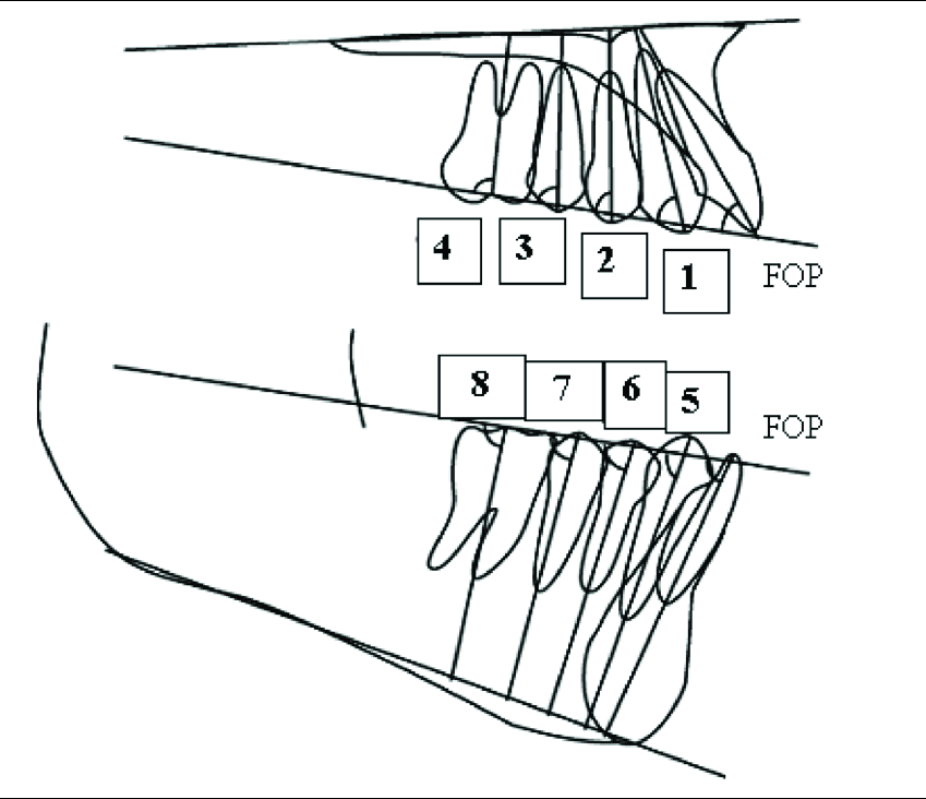
- Measurement of the axial angulations of the lateral teeth relative to the functional occlusal plane. (1) Maxillary canine; (2) maxillary first premolar; (3) maxillary second premolar; (4) maxillary first molar; (5) mandibular canine; (6) mandibular first premolar; (7) mandibular second premolar; (8) mandibular first molar

- Analyzed cephalometric variables. (1) FH-FOP angle; (2) SN-MP angle; (3) FMA; (4) gonial angle; (5) Y-axis. FH, Frankfort horizontal plane; FOP, functional occlusal plane; MP, mandibular plane; FMA, Frankfort-mandibular plane angle; Y-axis, angle between FH and S-Gn
Statistical analysis
Measurement error was determined by duplicate measurements of all the variables in a 1-month interval. The paired t-test was used to compare the intraobserver differences; a two-tailed P < 0.05 was regarded as significant in this analysis. The t-test was used to compare the mesiodistal angulation between maxilla and mandible. The mean values in the axial angulations were compared by repeated measures analysis of variance followed by Scheffe’s test among lateral teeth. To examine the correlations among the axial angulations, Pearson’s correlation was employed. P < 0.05 was regarded as critically significant in these analyses.
RESULTS
The intraobserver variation in the measurements was considered very small when compared with the measurement error (P < 0.01).
Five cephalometric variables are shown in Table 1. They are in normal range in Japanese standards. As shown in Table 2, the mean axial angulations of the maxillary canine, first premolar, second premolar, and first molar were 66.2°, 77.9°, 85.1°, and 89.4°, respectively. All the values showed statistical significance among maxillary teeth. On the other hand, the mean axial angulations of the mandibular canine, first premolar, second premolar, and first molar were 77.3°, 85.2°, 85.4°, and 84.4°, respectively. The axial angulation of canine was significantly smaller than premolars and molar in the mandible. No significant differences in the axial angulation of the second premolars. Noteworthily, the first molar values were approximately 90° relative to the FOP.
| Group | Mean±SD, degrees | ||||
|---|---|---|---|---|---|
| FH-FOP | SN-MP | FMA | Gonial angle | Y-axis | |
| Control | 10.9±2.5 | 33.5±5.6 | 26.4±5.6 | 118.5±10.5 | 63.5±3.4 |
| Japanese Norm | 11.6±4.2 | 34.0±3.9 | 28.5±3.9 | 122.1±6.4 | 62.9±2.7 |
SD – Standard deviation; FH – Frankfort horizontal; FOP – Functional occlusal plane
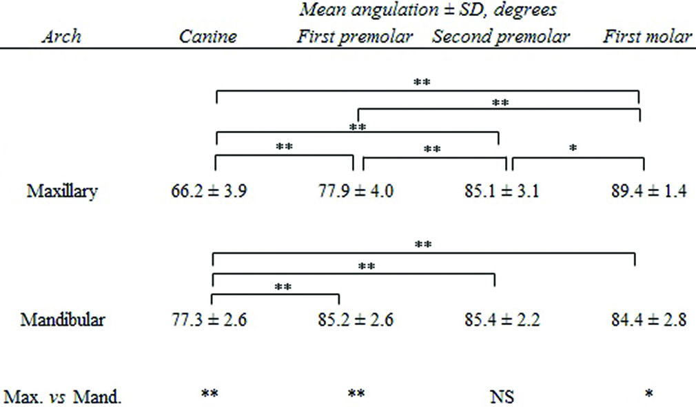 |
In addition, significant positive correlations (0.50–0.65) of the axial angulations were found with the canine, first premolar, and second premolar [Table 3].
| Tooth | Canine | First premolar | Second premolar | First molar |
|---|---|---|---|---|
| Canine | 1 | 0.64** | 0.5** | −0.22 |
| First premolar | 0.64** | 1 | 0.65** | −0.10 |
| Second premolar | 0.5** | 0.65** | 1 | −0.11 |
| First molar | −0.22 | −0.10 | −0.11 | 1 |
DISCUSSION
The present study was conducted to elucidate the mesial axial angulation of the maxillary and mandibular lateral teeth and the FH-FOP angle in the normal occlusions by cephalometric analysis. Significant differences in the axial angulations were noted between the maxillary and mandibular dentitions as previously shown by model analysis.[5]
The FOP was used as a reference plane to estimate the axial angulations in the present study. This plane may offer more advantages for analysis because the conventional occlusal plane is easily influenced by the vertical position of the incisors. Jacobson[7] concluded that a representative FOP would be a more appropriate plane for craniofacial analysis.
Progressive mesial tipping of the maxillary lateral teeth was noted. This tendency was more prominent in the first premolar than in the second premolar, because the first premolar is not prevented from tipping mesially before the eruption of the canine. In addition, the axial angulations were significantly correlated to each other. It may be explained in part by a fact that the angle of mesial angulation of erupting maxillary premolar relative to reference plane[8] on panoramic X-ray films showed the same results in the growing patients with mixed dentitions used as the subjects in the previous report.
Presumably, some factor caused mesial tipping of the lateral teeth germs in the alveolar bone. The underlying mechanism may reasonably be assumed as follows: The first molar erupts toward the end of the deciduous dentition, at around 6 years of age, and then, the deciduous teeth are replaced by the permanent teeth in the mixed dentition. While the first molar roots are forming and completing calcification, at around 6 and 9 years of age, respectively, the first and second premolar germs are close to the first molar and located at the same level as the first molar roots in the maxillary mixed dentition.[9] Hanai[10] reported that the arrangement of the teeth germs from the canine to the second molar straightens labiolingually and the second premolar germ descends to the level of the first premolar germ, although the canine germ is still in the highest position in the upper half of the maxillary process during the mixed dentition. Therefore, the axial angulation of the maxillary lateral teeth progressively increases in the mesial direction during the erupting stage.
In addition, the axes of the maxillary teeth tend to converge in the maxilla, whereas the opposite is true in the mandible.[11] In general, the maxillary lateral teeth are angulated more mesially than the mandibular ones.[12,13] Therefore, maxillary anterior crowding with high canines and slight mandibular incisor crowding may involve completely different mechanisms; however, the cause of this malocclusion has not been fully elucidated. One factor may be the prominent mesial axial angulation of the maxillary lateral teeth relative to the FOP.[5] Such finding may explain why crowded maxillary lateral teeth germs are encountered frequently during panoramic radiograph analysis.
Further, the first molar is located perpendicular to the FOP in most patients. The reason may be that the first molar is the principal tooth supporting the bite force.[14,15] For mechanically beneficial occlusion, the maxillary first molar should be perpendicular to the FOP.
This study has some limitations due to the sample size being relatively small, and while this method is established to compare data easily, cephalometric analysis provides only two-dimensional data, therefore, is not as reliable as a three-dimensional (3D) diagnostic tool. It is thus anticipated hopefully to use 3D imaging techniques,[16,17] which provide additional detail information about the positional relationship between the first molar root and the lateral teeth germs, in the normal and crowding cases.
CONCLUSIONS
The maxillary lateral teeth are angulated more mesially than the mandibular ones relative to the FOP. Moreover, progressive mesial tipping of the maxillary lateral teeth was found, and the axial angulations were significantly correlated to each other although the mandibular premolars and molar are angulated similarly.
Financial support and sponsorship
Nil.
Conflicts of interest
There are no conflicts of interest.
References
- Theoretical and practical aspects of crowding in the human dentition. J Am Dent Assoc. 1974;89:139-53.
- [Google Scholar]
- Facial and dental arch morphology in children with and without early loss of deciduous molars. Am J Orthod. 1978;73:47-58.
- [Google Scholar]
- An examination of dental crowding and its relationship to tooth size and arch dimension. Am J Orthod. 1983;83:363-73.
- [Google Scholar]
- Orthodontics: Current Principles and Techniques (2nd ed). St. Louis, MO: CV Mosby Co.; 1994. p. :321-7.
- Changes in the crown angulation and dental arch widths after nonextraction orthodontic treatment: Model analysis of mild crowding with high canines. Open J Stomatol. 2012;2:188-94.
- [Google Scholar]
- A study on the tooth material in Japanese adults of normal occlusion, its relationship to control and basal arches. J Jpn Orthod Soc. 1957;16:36-46.
- [Google Scholar]
- Change in themesiodistal axial inclination of the maxillary lateral teeth during the mixed dentition stage: Morphometric analysis of panoramic radiographs from two cases of mild crowding with a high canine. APOS Trends Orthod. 2016;6:24-30.
- [Google Scholar]
- Prediction of mandibular incisor and canine crowding changes in the mixed dentition. Am J Orthod. 1985;88:47-63.
- [Google Scholar]
- Studies on the movement and the growth of the permanent tooth germs in the maxilla of the children with the eruption of the permanent teeth. Shikwa Gakuho. 1976;76:1351-412.
- [Google Scholar]
- Arrangement in the jaws of the roots of the teeth. J Am Dent Assoc. 1963;67:779-97.
- [Google Scholar]
- A normative study to evaluate inclination and angulation of teeth in North Indian population and comparision of expression of torque in preadjusted appliances. J Orthod Sci. 2014;3:81-8.
- [Google Scholar]
- Occlusal force distribution on the dental arch during various levels of clenching. J Oral Rehabil. 1999;26:932-5.
- [Google Scholar]
- Changes in occlusal force and occlusal contact area after active orthodontic treatment: A pilot study using pressure-sensitive sheets. J Oral Rehabil. 2002;29:484-91.
- [Google Scholar]
- Mesiodistal angulation and faciolingual inclination of each whole tooth in 3-dimensional space in patients with near-normal occlusion. Am J Orthod Dentofacial Orthop. 2012;141:604-17.
- [Google Scholar]
- A new method to measure mesiodistal angulation and faciolingual inclination of each whole tooth with volumetric cone-beam computed tomography images. Am J Orthod Dentofacial Orthop. 2012;142:133-43.
- [Google Scholar]


